
- Discussion:
- pathoanatomy:
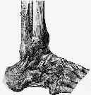
- fibular collateral ligament is made up of three separate structures;
- these are not as strong as medial ligaments, since lateral support of ankle is also provide by the fibula;
- anterior talofibular ligament
- calcaneofibular ligament
- posterior talofibular Ligaments
- cervical ligaments:
- ref: Elongation behavior of calcaneofibular and cervical ligaments during inversion loads applied in an open kinetic chain.
- deltoid ligament resists abduction & lateral rotation of foot;
- sudden and forceful eversion, inverson, or rotation of the foot may tear the ligament stressed, or by acting through the ligament, may avulse the attached malleolus;
- in the vast majority of cases there will be a tear thru the ligament mid-substance;
- associated talar compression of opposite malleolus may cause frx;
- anterior talofibular ligament:
- w/ inversion force of foot, there is injury to anterolateral capsule, ATFL, & anterior tibiofibular ligament;
- about 40% of patients will have this injury type;
- calcaneofibular: as force progresses, this ligament is injured as well;
- in about 58% of cases, there will be a tear of both the ATFL and the CF ligament;
- finally in a small number of cases (3%) there will be tears of the above two ligament and the posterior talofibular ligaments;
- differential diagnosis: ankle sprains:
- anterior impingement snydrome of the ankle
- calcaneocuboid joint injuries;
- type of inversion sprain that involves a portion of ligaments overlying the calcaneocuboid joint;
- causes immediate severe disability with pain, swelling, and tenderness that is localized to the region of the joint;
- frx of anterior process of calcaneus
- frx of the lateral talar processes
- frx of the posterior process
- syndesmotic sprain (high ankle sprain)
- midtarsal frx
- subtalar joint sprain
- osteochondral lesions of talus
- peroneal tendon disruption
- tarsal coaliltion
- Physical Exam:
- be sure to look for concomitant syndesmotic and subtalar instability;
- note any dysesthesia from the peroneal nerves, and point this out to the patient;
- Radiographic Studies for Ankle Sprains:
- tibiotalar tilt
- Non Operative Treatment:
- non operative treatment is indicated for the majority of severe ankle sprains, and most patients w/ significant sprains will be back to work with in 10 days;
- prognosis (from Gerber, et al (1998))
- factor most predictive of delayed recovery and residual symptoms is concomitant syndesmotic sprain;
- most patients will be back to playing sports at 6 weeks, but about 1/2 will continue to significant symptoms even at 6 months;
- prognosis of tibiotalar tilt:
- despite the fact that most severe ankle sprains will initially demonstrate 15-18 deg of tibio-talar tilt (and in some cases will be as large as 40 deg), in most
cases the ankle will stabilize down to 6 deg with conservative treatment;
- in patients w/ less than 15 deg of initial tibiotalar tilt, approximately 13% will have a poor result;
- in patients w/ more than 15 deg of initial tibiotalar tilt, approximately 22% will have a poor result;
- references:
- Management and rehabilitation of ligamentous injuries to the ankle.
- Radiological and muscular status following injury to the lateral ligaments of the ankle. Follow-up of 144 patients treated conservatively.
- Clinical and social status following injury to the lateral ligaments of the ankle. Follow-up of 144 patients treated conservatively.
- Persistent disability associated with ankle sprains: A prospective examination of an athletic population.
- Functional properties of adhesive ankle taping: Neuromuscular and mechanical effects before and after exercise.
- Spraining the Ankle Without Straining Credulity
- I Thought Everyone Knew That RICE Is Effective in Treating Acute Ankle Sprains!
- Operative Management:
- indications:
- in the meta-analysis report by Pijnenburg, et al, the authors concluded that a no-treatment strategy for ruptures of the lateral ankle ligaments leads to more residual symptoms.
- operative treatment leads to better results than functional treatment, and functional treatment leads to better results than cast immobilization for six weeks;
- reference:
- Treatment of Ruptures of the Lateral Ankle Ligaments: A Meta-Analysis.
- prior to considerations for surgery, ensure that subtalar instability is not present;
- Modified Brostrom Procedure:
- generally operative treatment is considered when there is a persistant tibiotalar tilt greater than 20 deg;
- EDB transfer for chronic lateral ankle instability:
- EDB is detached proximally from the calcaneus and is sutured to the periosteum of the fibula;
- procedure helps to restore proprioception to the ankle;
- associated injuries which may require treatment:
- in report by DiGiovanni, et al (2000), the authors sought to determine injuries associated with lateral instability at the time of surgery;
- 61 patients underwent a primary ankle lateral ligament reconstruction for chronic instability between 1989 and 1996;
- in addition to the ligament reconstruction, all patients had evaluation of the peroneal retinaculum, peroneal tendon inspection by
routine opening of the tendon sheath, and ankle joint inspection by arthrotomy;
- at surgery no patients were found to have isolated lateral ligament injury;
- injuries found most often by direct inspection included:
- peroneal tenosynovitis, 47/61 patients (77%);
- anterolateral impingement lesion, 41/61 (67%);
- attenuated peroneal retinaculum, 33/61 (54%);
- ankle synovitis, 30/61 (49%);
- intra-articular loose body, 16/61 (26%); peroneus brevis tear, 15/61 (25%);
- talus osteochondral lesion, 14/61 (23%); medial ankle tendon tenosynovitis, 3/61 (5%);
- Associated injuries found in chronic lateral ankle instability.
- peroneal tendon disruption
- peroneus brevis tear:
- longitudinal tears of the peroneus brevis are associated w/ ankle sprains;
- look for tendon tear at the level of the distal fibula;
- persistent swelling along the peroneal tendon sheath is a reliable sign for peroneus brevis tendon tear;
- this injury tends to occur from peroneal tendon subluxation over the posterolateral edge of the fibula;
- inciting cause is incompetence of the superior peroneal retinaculum;
- this allows subluxation of the peroneal tendons and mechanical attrition of the peroneus brevis tendon against the posterior ridge of the fibula;
- treatment: needs to address the tear and the peroneal subluxation;
- w/ damage of less than 50% tendon substance, consider tendon debridment;
- w/ damage of more than 50% of the tendon cross sectional area, consider excision of the damaged segment and tenodesis to the peroneus longus;
- valgus osteotomy:
- Idiopathic cavovarus and lateral ankle instability: Recognition and treatment implications relating to ankle arthritis.
- references:
- Ruptures of the fibular collateral ligaments of the ankle. Result study of immediate surgical treatment.
- Static or dynamic repair of chronic lateral ankle instability. A prospective randomized study.
- Reconstruction of the lateral ligaments of the ankle for chronic lateral instability.
- Reconstruction of the lateral ligaments of the ankle using the plantaris tendon.
- Long-term results of the Chrisman-Snook operation for reconstruction of the lateral ligaments of the ankle.
- Lateral ligament reconstruction of the ankle with a modified Watson-Jones operation.
- Results of Watson-Jones ankle reconstruction for instability. The influence of articular damage.
- A new operation for chronic lateral ankle instability.
- Secondary reconstruction of the lateral ligaments of the ankle.
- Long-term results of the Evans procedure for lateral instability of the ankle.
- The effect of musculus extensor digitorum brevis transfer for chronic lateral ankle instability.
- Associated injuries found in chronic lateral ankle instability.
Treatment for acute tears of the lateral ligaments of the ankle. Operation, cast, or early controlled mobilization.
The contribution of the anterior talofibular ligament to ankle laxity.
Talar shift. The stabilizing role of the medial, lateral, and posterior ankle structures.
The deltoid ligament. An evaluation of need for surgical repair.
Injury to the lateral ligaments of the ankle.
The influence of dorsiflexion in the treatment of severe ankle sprains: an anatomical study.
Instability of the ankle after injury to the lateral ligament.
Early and late repair of lateral ligament of the ankle.
Year Book: A Prospective Study of the Treatment of Severe Tears of the Lateral Ligament of the Ankle.
Stability of the loaded ankle: Relation betweeen articular restraint and primary and secondary static restraints.
Treatment of Ruptures of the Lateral Ankle Ligaments: A Meta-Analysis.
Long-term results after modified Brostrom procedure without calcaneofibular ligament reconstruction