
- Mechanism: ACL Tear:

- Physical Exam of ACL Tears: (see knee exam)
- Anterior Drawer Test
- Anteromedial Rotatory Stability:
- Anterolateral Rotatory Instability
- Clunk Test
- Lachman
- Losee Test
- Partial ACL
- Pivot shift
- Reverse Pivot Shift Test
- Hemarthrosis:
- greater than70% of pts w/ acute hemarthrosis will have ACL tear;
- severe swelling of the knee typically develops within two hours of injury because of hemarthrosis;
- hemarthrosis will develop over 6-24 hours;
- if effusion develops immediately after injury, one should suspect an osteochondral fracture;
- presence or absence of fat in aspirated fluid is key distinction;
- Discussion:
- ensure that patient has a FROM and no indication of arthrofibrosis:
- pivot shift with a positive Lachman's;
- combined injuries:
- posterior laxity;
- MCL instability:
- in most cases, the torn MCL will go on to heal if treated conservatively;
- ACL reconstruction should be delayed until medial laxity is not present, otherwise the combined instability may comprimise the final result;
- posterolateral instability:
- should be managed operatively along with the ACL reconstruction;
- neglected posterolateral instability may lead to ACL reconstruction failures;
- ref: Does Physiologic Posterolateral Laxity Influence Clinical Outcomes of Anterior Cruciate Ligament Reconstruction?
- meniscal tear:
- strongly consider repair at the time of ACL reconstruction;
- pass sutures prior to the ACL reconstruction but delay tying sutures down until the reconstruction is completed;
- references:
- Treatment of acute isolated and combined ruptures of the anterior cruciate ligament. A long term follow up study.
- Decreased range of motion following acute versus chronic anterior cruciate ligament reconstruction.
- Diff Dx:
- consider quadriceps or patella tendon rupture;
- PCL ruptures may give a "false positive" Lachman test;
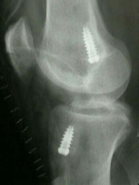
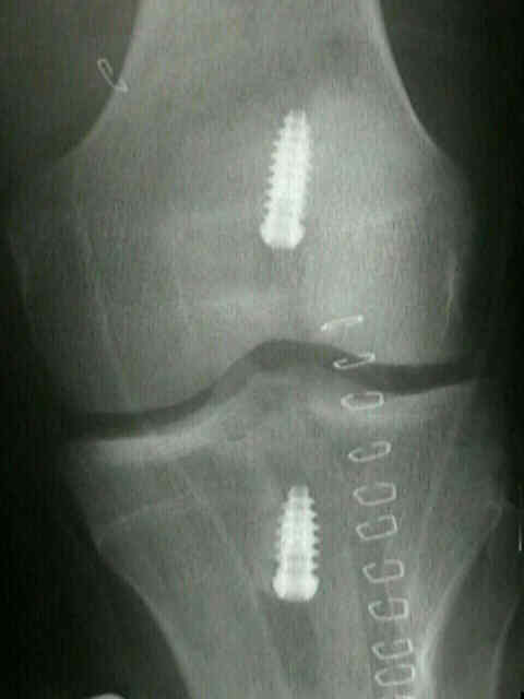
- Radiographs:
- hyper-extension lateral view allows assessment of slope of intercondylar roof in relation to the tibial plateau;
- this may help with placement of the tibial tunnel (helps avoid graft impingement);
- Segond fracture
- small avulsion frx of lateral tibial condyle just below joint line is now recognized as a sign of injury of ACL;
- small avulsion frx of proximal part of the tibia that is seen just proximal to fibular head on the anteroposterior roentgenogram, is
nearly always associated w/ torn ACL;
- references:
- The Segond Fracture: A Bony Injury of the Anterolateral Ligament of the Knee
- The Segond fracture of the proximal tibia: A small avulsion that reflects major ligamentous damage.
- references:
- Patella baja in anterior cruciate ligament reconstruction of the knee.
- Lateral capsular sign: x-ray clue to a significant knee instability.
- Relation of the fibular head sign to other signs of anterior cruciate ligament insufficiency. A follow-up letter to the editor.
- Patellar tendon graft reconstruction for midsubstance ACL rupture in junior high school athletes. An algorithm for management.
- Assessment of patellar height after autogenous patellar tendon anterior cruciate ligament reconstruction.
- Fracture of the posterior aspect of the lateral tibial plateau: radiographic sign of anterior cruciate ligament tear.
- Does Posterior Tibial Slope Influence Knee Functionality in ACL-Deficient and ACL-Reconstructed Knee?
- Anatomic Graft Placement in ACL Surgery: Plain Radiographs Are All We Need
- Posterior Tibial Slope Influences Static Anterior Tibial Translation in Anterior Cruciate Ligament Reconstruction
A Minimum 2-Year Follow-up Study
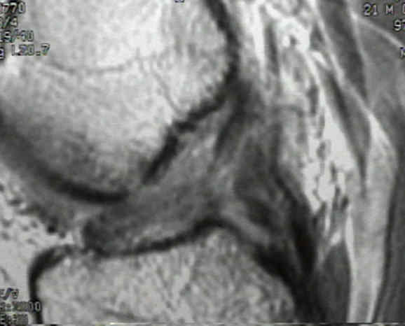
- MRI of Knee:
- normal anatomy:
- distal ligament may show a striated signal caused by interspersed fat and synovium between the 2 bundles;
- proximal ligament appears dark
- any discontinuity or signal change in the ligament is indicative of ACL tear;
- indirect signs of ACL tear:
- always look for signs of additional injury (meniscal tear, PCL tear, LCL tear);
- femoral osteochondral lesions and/or tibial plateau bone bruises may diminish the eventual postoperative result;
- look for increase in signal on T2-weighted images and decreased signal on T1-weighted images
- often there will be focal areas of increased signal in the posterior aspect of lateral tibial plateau and mid portion of the lateral femoral condyle.
- signal changes occurs as a consequence of pivot shift injury:
- combination of signal changes in lateral femoral condyle and posteror lateral tibial plateau results from of a valgus-
external rotation of the femur on the fixed tibia;
- abnormal slope of ACL;
- avulsion of the anterior tibial spine;
- segond fracture: capsular avulsion fracture of the lateral tibial plateau;
- kissing contusions involving the anterior tibia and femur resulting from hyperextension injury;
- references:
- The accuracy of selective magnetic resonance imaging compared with the findings of arthroscopy of the knee.
- "Bone Bruises" on magnetic resonance imaging evaluation of anterior cruciate ligament injuries.
- Occult posttraumatic osteochondral lesions of the knee: prevalence, classification, and short-term sequelae evaluated with MR imaging
- Associated Findings:
- varus knee:
- whereas a varus knee probably does not cause ACL tear, it is well known that a varus knee will have a negative influence on both operative and non-op treatment;
- w/ signficant varus deformity consider high tibial osteotomy prior to ACL reconstruction;
- references:
- High tibial osteotomy and ligament reconstruction in varus angulated, anterior cruciate ligament-deficient knees: a 2-7 year follow up study.
- osteochondral defects
- meniscal tear:
- menisci may become trapped between femoral and tibial condyles.
- vertical longitudinal & " bucket-handle tear;"
- as tibia subluxates anteriorly, the posterior horns of menisci become trapped between articular surfaces of femur and tibia;
- if torn meniscus fragment remains forward of the femoral condyle when the tibia reduces, knee is "locked;"
- note that the combination of ACL tear and meniscal tear is especially likely to contribute to instability, and early osteo-arthritis;
- ACL tears combined w/ medial meniscal tears result in more anterior translation than lateral meniscal tears;
- management:
- Shelbourne KD and Johnson GE (1993), advocate staged treatment of concomitant bucket hand and ACL injuries;
- theoretical advantages of staged repair and reconstruction:
- more aggressive use of repair rather than removal of a displaced torn meniscus;
- prevention of problems in regaining range of motion;
- allows a second look to judge the success of meniscal repair;
- allows time for the patient to prepare for ACL reconstruction;
- references:
- Locked bucket-handle meniscal tears in knees with chronic anterior cruciate ligament deficiency.
- MCL tear:
- treated non operatively, most MCL tears will heal with non operative treatment;
- some surgeons will delay ACL reconstruction until there is near complete resolution of medial instability;
- references:
- The treatment of acute combined ruptures of the anterior cruciate and medial ligaments of the knee.
- Reconstruction of ACL alone in the treatment of a combined instability with complete rupture of MCL. A prospective study.
- posterolateral instability:
- these patients may demonstrate an asymmetric recurvatum deformity, which is most pronounced during gait;
- exam: external rotation recurvatum test and reverse pivot shift test
- LCL instability should be managed concurrently w/ ACL reconstruction;
- it is important to distinguish this type of instability from one plane posterior instability;
- isolated PCL/ACL reconstruction will correct the one plane instability but will not correct the rotatory instability;
- in fact, ACL reconstruction may actually increase the patient's sensation of rotatory instability;
- reference:
- Treatment or acute and chronic combined anterior cruciate ligament and posterolateral knee ligament injuries.
- incomplete knee extension:
- most often is a result of a "cyclops lesion," which is due to a stum
Original Text by Clifford R. Wheeless, III, MD.
Last updated by Clifford R. Wheeless, III, MD on Sunday, April 26, 2015 3:06 pm