
- See: TKR Menu: (Function and Activity after TKR)
- Natural History of Painful TKR:
- references:
- Pain and Depression Influence Outcome 5 Years after Knee Replacement Surgery.
- Impact of Psychological Distress on Pain and Function Following Knee Arthroplasty
- Potential Causes of TKR failure:
- early failures:
- references:
- Early failures in total knee arthroplasty.
- Early failures among 7,174 primary total knee replacements: a follow-up study from the Norwegian Arthroplasty Register 1994-2000.
- infected TKR
- bone scans:
- may indicate loosening after 6-12 months, but can not distinguish between septic and aseptic loosening;
- aspiration: for gm stain & culture:
- most accurate method of dx, and is required prior to all revisions;
- remember that knee aspiration can yield false positives up to 25% of the time;
- sed rate and CRP
- Use of erythrocyte sedimentation rate and C-reactive protein level to diagnose infection before revision total knee arthroplasty. A prospective evaluation.
- component misplacement:
- varus malalignment has no influence on clinical outcome in midterm follow-up after total knee replacement
- rotation of the femoral component:
- references:
- Rotational malalignment of the femoral component in total knee arthroplasty.
- [Rotational malalignment of the components may cause chronic pain or early failure in total knee arthroplasty.]
- rotation of tibial component:
- references:
- Internal rotational error of the tibial component is a major cause of pain after total knee replacement
- mis-sizing of components
- medial tibial overhang;
- over-sizing the femoral component;
- references:
- Overhang of the femoral component in total knee arthroplasty: risk factors and clinical consequences.
- Unique relationship between osteophyte and femoral-tibia component size mismatch in determining polyethylene wear in primary total knee arthroplasty: a case report.
- The influence of the implant size on the outcome of unconstrained total knee arthroplasty.
- knee arthroplasty instability:
- remnant medial meniscus:
- An impinging remnant meniscus causing early polyethylene failure in total knee arthroplasty: a case report.
- Pseudomeniscal Synovial Impingement After Unicondylar Knee Arthroplasty
- patellar problems:
- patellar frx
- patellar clunk syndrome
- patellar subluxation
- eccentric patellar button placement (causing patellar tilt and contact between the lateral patellar facet and the lateral femoral condyle);
- asymmetric patellar resurfacing: tendency to underresface the medial patellar facet;
- ref: The John Insall Award: control-matched evaluation of painful patellar Crepitus after total knee arthroplasty.
- painful tibial stem
- Tibia pain at end of stem with stemmed revision total knee arthroplasty: treatment with cortical strut graft technique.
- Tibial stem tip pain in stemmed revision total knee arthroplasty: treatment with tension band plating.
- malalignment of feet:
- Persistent hindfoot valgus causes lateral deviation of weightbearing axis after total knee arthroplasty.
- Evaluation of knee and hindfoot alignment before and after total knee arthroplasty: a prospective analysis.
- component failure:
- fibrous ingrowth:
- pain resulting from fibrous ingrowth should always be considered in a patient with a press fit femoral or tibial component;
- polyethylene failure:
- look for asymmetrical polyethylene thickness;
- polyethylene may wear out, especially postero-medially;
- may be associated w/ osteolysis;
- polyethylene wear may yield green fluid aspirate;
- tibial component frx
- patellar frx
- ref: Mechanisms of failure of the femoral and tibial components in total knee arthroplasty.
- neuropathic pain:
- back pain:
- Patient-reported outcomes after total knee replacement vary on the basis of preoperative coexisting disease in the lumbar spine and other nonoperatively treated joints: the need for a musculoskeletal comorbidity index.
- The natural history of pain and neuropathic pain after knee replacement: a prospective cohort study of the point prevalence of pain and neuropathic pain to a minimum three-year follow-up.
- neuroma: (saphenous nerve)
- Neuroma of the infrapatellar branch of the saphenous nerve a cause of reversible knee stiffness after total knee arthroplasty.
- Fate of the infrapatellar branch of the saphenous nerve post total knee arthroplasty
- Infrapatellar Saphenous Neuralgia After TKA Can Be Improved With Ultrasound-guided Local Treatments
- gout / pseudogout as a cause of painful TKR (gout and pseudogout):
- allergy:
- Pain in a chromium-allergic patient with total knee arthroplasty: disappearance of symptoms after revision with a special surface-coated TKA — a case report
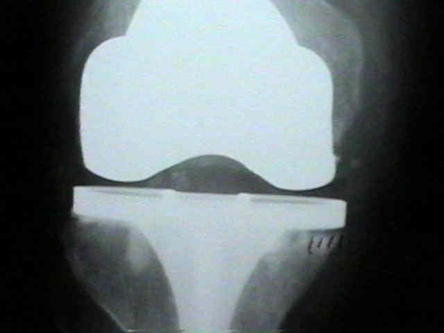
- Radiographs:
- comparing sunrise views and lateral films to the native knee (or ipsilateral pre-op films) can help clarify patellofemoral problems
(tracking, "stuffed" patellofemoral joint, lateral facet contact due to an undersized component, etc.)
- component loosening:
- loosening w/ knee replacements is most often due to subsidence, in which component actually sinks into bone;
- malalignment, esp. varus alignment, usually has a causal relationship to this failure mode;
- when its unclear whether loosening or slight subsidence is present, consider obtaining radiographs under flouroscopic control;
- also consider need for subtraction arthrogram inorder to emphasize the space between bone and cement or between cement and prosthesis;
- also indicated for sublte femoral component loosening, which is difficult to diagnose on plain radiographs;
- in study by Vyskocil et al, (1999), authors found that flourscopically assisted xrays identified significantly more radioluncent
lines in femoral components than were found by conventional xrays;
- authors note that deviation of x-ray beam of only 2.3 deg to component interface could obscure a radiolucent line of 2 mm, but despite this,
authors were unable to demonstrate a significant advantage to flouroscopically assisted radiographs to detect tibial loosening;
- references:
- Radioluncent lines and component stability in knee arthroplasty. Standard versus flouroscopically assisted radiographs.
- Fluoroscopic evaluation of the painful total knee arthroplasty.
- Radionuclide imaging of asymptomatic versus symptomatic total knee arthroplasties.
- Early Femoral Component Loosening of Constrained Condylar Primary Total Knee Arthroplasties Inserted Without Stems
- ACR Appropriateness Criteria® imaging after total knee arthroplasty.
- Assessment of radionuclide arthrography in the evaluation of loosening of knee prostheses
- Treatment Options:
- Role of Arthroscopy following TKR
- Revision TKR
- Physical Therapy following TKR
Intraarticular botulinum toxin a for refractory painful total knee arthroplasty: a randomized controlled trial.
Recurrent hemarthrosis after knee joint arthroplasty: etiology and treatment