
- See: differential diagnosis of posterior fossa masses 
- Discussion:
- popliteal artery is the continuation of superficial femoral artery at hiatus of the adductor magnus muscle;
- artery is anchored proximally by tendinous insertion of adductor magnus upon the medial femoral epicondyle;
- it runs posterior to the distal femur, behind knee joint;
- at the supracondylar ridge, the artery gives off the blood supply to the knee;
- just above the knee joint, the following arteries are given off:
- medial and lateral sural arteries
- cutaneous branch that acompanies the small saphenous vein; 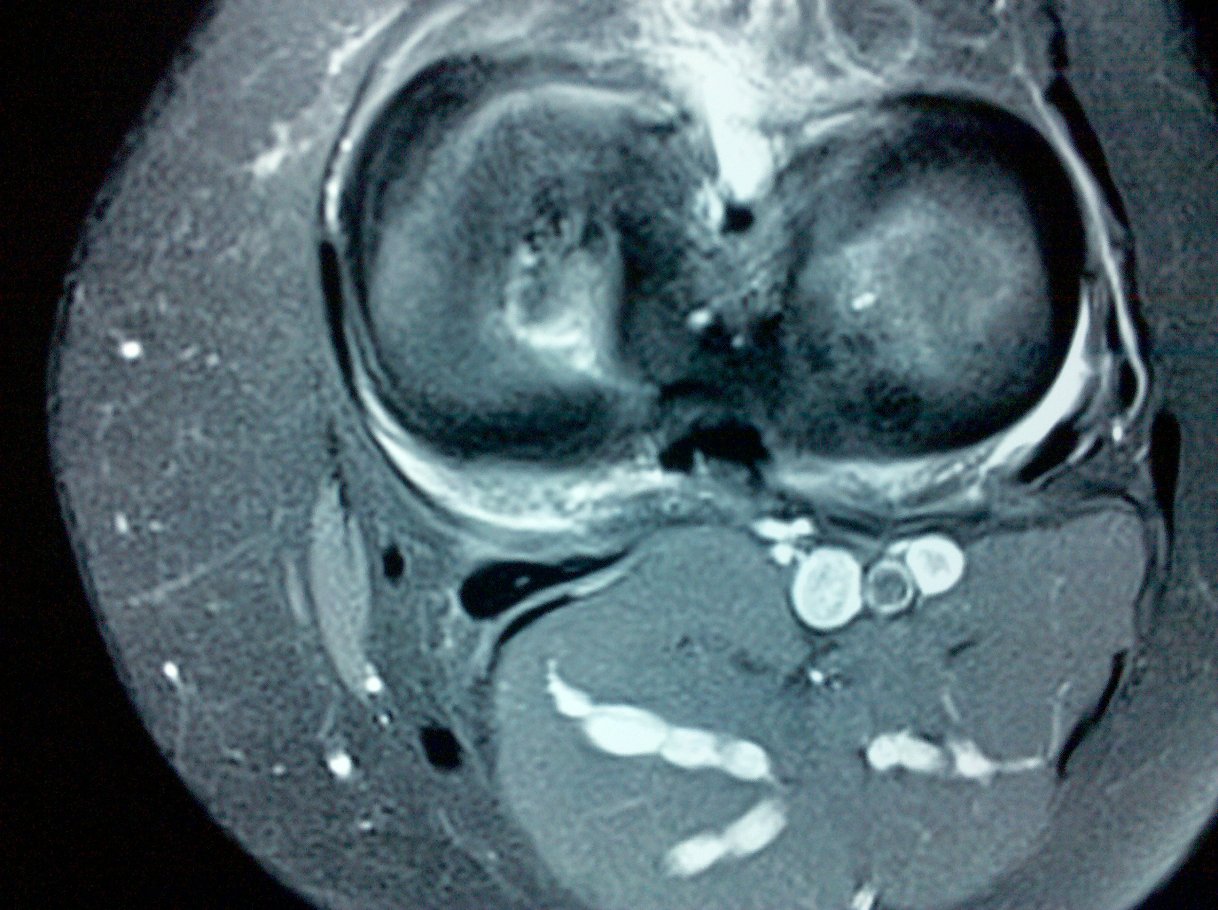
- middle genicular artery;
- anatomy at the knee:
- at the level of the knee joint, the popliteal artery gives off the medial and lateral genicular arteries;
- popliteal artery lies behind posterior horn of the lateral meniscus;
- only thin layer of fat separates popliteal artery from thin posterior capsule behind posterior horn of lateral meniscus;
- popliteal artery lies anterior to popliteal vein and 9 mm posterior to the posterior aspect of tibial plateau in 90
degrees of flexion;
- distally the femoral artery is fixed by tendon of soleus as it descends from its insertion on the medial aspect
of tibial plateau;
- before passing deep to fibrous arch over soleus muscle, it divides into anterior and posterior tibial arteries 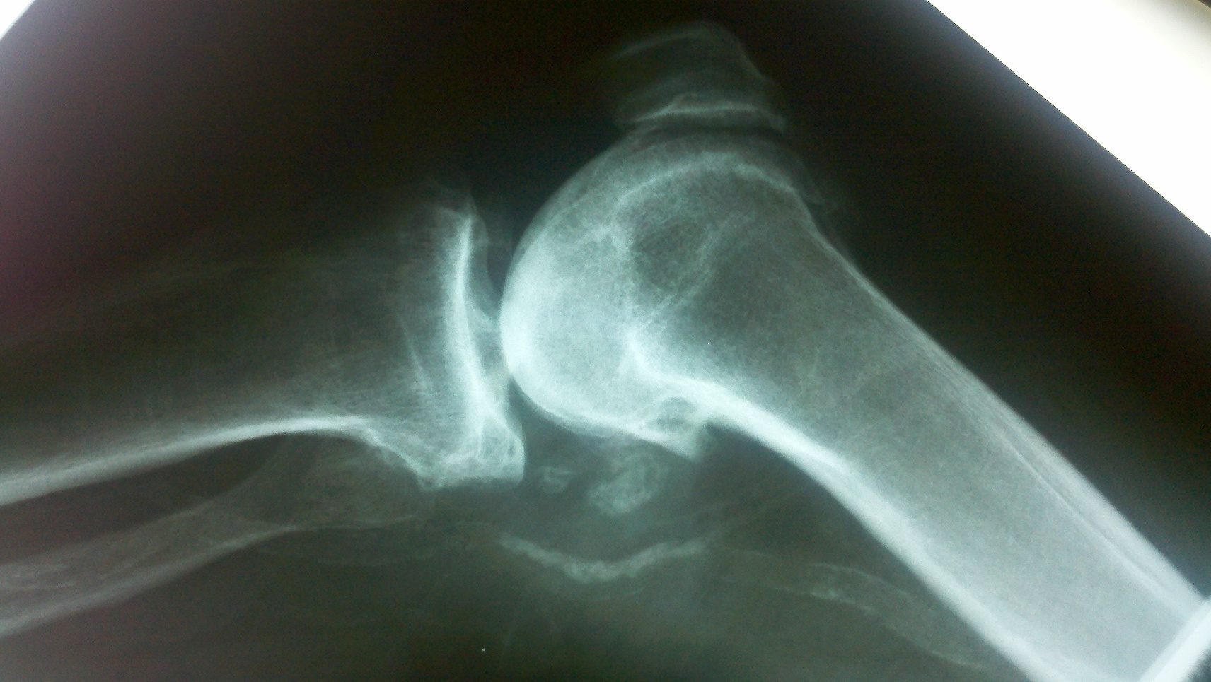
at distal aspect of popliteus muscle;
- popliteal artery normally branches into anterior tibial artery & tibioperoneal trunk at distal border of popliteus muscle;
- references:
- Popliteal artery injury complicating arthroscopic menisectomy.
- Postoperative aneurysm of the popliteal artery after arthroscopic meniscectomy.
- Pseudoaneurysm of the popliteal artery following arthroscopic meniscectomy.
- The effect of knee flexion on the popliteal artery and its surgical significance.
- Proximity of the posterior cruciate ligament insertion to the popliteal artery as a function of the knee flexion angle: implications for posterior cruciate ligament reconstruction.
- Prevalence and surgical significance of a high-origin anterior tibial artery
- Delayed Pseudoaneurysm of the Popliteal Artery Following ACL Reconstruction
.............................................................................................................................................................................................................................................................................................................................
- Damage from Knee Dislocation: (see knee dislocation)
- see management: and popliteal vessel disruption from knee dislocation:
- anterior dislocation:
- hyperextension causes popliteal artery to be stretched;
- pt typically suffers intimal separation over long segment;
- posterior dislocation:
- less common than anterior dislocation;
- less common, due to even greater forces needed to overcome strength of extensor muscles of leg;
- popliteal artery usually suffers direct contusion or intimal fracture;
- references:
- Arterial injuries associated with complete dislocation of the knee.
- Arterial injury complicating knee disruption.
- Surgical Approach - Medial Incision:
- above knee portion of popliteal artery is exposed thru medial thigh incision used for exposure of the saphenous vein;
- incision is made over and parallel to the sartorius muscle;
- deep fascia of thigh is incised along lateral (superior) margin of sartorius;
- for exploration of the artery, it is advantageous to retract the sartorius, semitendinous, and gracilis anteriorly;
- for this reason, proximal portion of incision runs the length distal third of thigh, parallels patella, & then runs 1 cm posterior to posterior border of tibia;
- popliteal artery is identified where it exits adductor canal;
- dissection carried distally, in plane of arterial adventia;
- below knee, popliteal artery is exposed thru a medial calf incision (vein harvest incision) approx one fingers breadth below the tibia margin;
- after incision of deep fascia, medial head of gastrocnemius is retracted inferiorly;
- artery is found slightly medial to popliteal vein;
- perform circumferential dissection & place Silastic vessel loop around artery, to allow arterial retraction;
- exposure of posterior tibial or peroneal arteries in calf by extending medial calf incision;
- division of medial of soleus origin on tibia allows exposure to vessels;
- posterior tibial artery is encountered first along with its paired tibial veins;
- Problems associated with Posterior Incision;
- it is difficult to extend the incision proximally or distally because of deep situation of the artery
- position of the patient makes it awkward to obtain saphenous vein graft from the thigh;
- Anastomosis:
- before the anastomosis is completed, a no 4 fogarty catheter is passed distally to remove clots, and the graft is flushed to remove clots;
- all anastomoses are performed with a continuous, everting suture of 6-0 polypropylene or 7-0 polytetrafluoroethylene (PTFE);
- Popliteal Emboli:
- can be removed thru common femoral arteriotomy.
- this approach also allows extraction of concomitant but clinically unsuspected emboli in the profunda femoris artery;
- complete clot removal from distal popliteal artery and trifurcation vessels, is complicated by tendency to pass directly into peroneal artery when passed from groin;
- following proximal and distal control a longitudinal arteriotomy is made in the distal popliteal artery, just opposite the takeoff of anterior tibial artery;
- 2 French Fogarty catheter is passed directly into ea tibial artery;
- after clot removal & distal flushing with heparinized saline arteriotomy is closed w/ running stitch of fine monofilament suture;
- completion angiography is necessary unless palpable pulses return;
- Popliteal Aneurysms:
- see: differential diagnosis of posterior fossa masses
- upto 50% are bilateral, and when it is bilateral, look for AAA;
- thrombotic complications of popliteal artery aneurysms are to be expected during conservative management of these lesions;
- majority may progress to complete occlusion, even when small;
- inaddition to thrombosis, risks include thrombophlebitis due to compression on popliteal vein, & pain from pressure on sural nerve;
- references:
- Pseudoaneurysm of the popliteal artery with an unusual arteriographic presentation. A case report.
- Popliteal arterial aneurysms. Their natural history and management.
- Popliteal artery aneurysms: tried, true, and new approaches to therapy.
- Popliteal aneurysm presenting as chronic exertional compartment syndrome.
- Popliteal Entrapment Syndrome:
- Popliteal vascular entrapment syndrome caused by a rare anomalous slip of the lateral head of the gastrocnemius muscle.
- Popliteal artery entrapment syndrome.
- References:
Advances in the management of acute popliteal vascular blunt injuries.
Lateral approach to the popliteal artery.
Scientific Papers: Successful Repair of Pediatric Popliteal Artery Trauma.
Successful management of trifurcation injuries.
Improved limb salvage in popliteal artery injuries.
Injury to the popliteal artery.
Injury to the popliteal vessels: the Lebanese war experience.
Knee salvage utilizing the myocutaneous principle.
Blunt popliteal artery injury with complete lower limb ischemia: is routine use of temporary intraluminal arterial shunt justified?
Illustrated Encyclopedia of Human Anatomic Variation: Popliteal Artery