
 - Discussion: Tibial Plateau Frx Menu
- consists of wedge frx of medial & lateral plateau;
- if articular depression is present, it is usually present on the lateral plateau;
- frx may have an inverted Y appearance, w/ the articular frx originating in the intercondylar region;
- associated injuries:
- 50% of plateau fractures will have peripheral meniscal detachment;
- ACL avulsions may occur in about 1/3 patients;
- consider possibility of spontaneously reduced knee dislocation;
- ref: Soft tissue injury of the knee after tibial plateau fractures.
- compartment sydrome
- popliteal artery injury
- referernces:
- Complications associated with internal fixation of high-energy bicondylar tibial plateau fractures utilizing a two-incision technique.
- Infection After Spanning External Fixation for High-Energy Tibial Plateau Fractures: Is Pin Site-Plate Overlap a Problem?
- Diagnosis and treatment of hyperextension bicondylar tibial plateau fractures
- Radiographs: 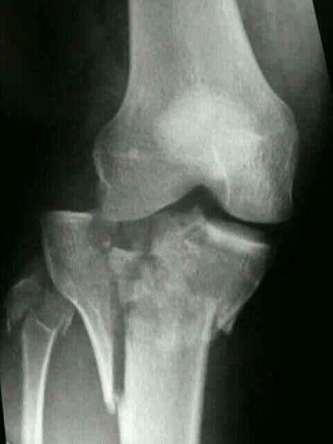
- Non Operative Treatment:
- because of soft tissue attachment to fracture fragments, traction occassionally provides an acceptable reduction & once frx has
become sticky may be managed in a cast brace;
- note, however, that loss of frx position & alignment is common when plaster cast is used after bicondylar fractures;
- PreOp Planning
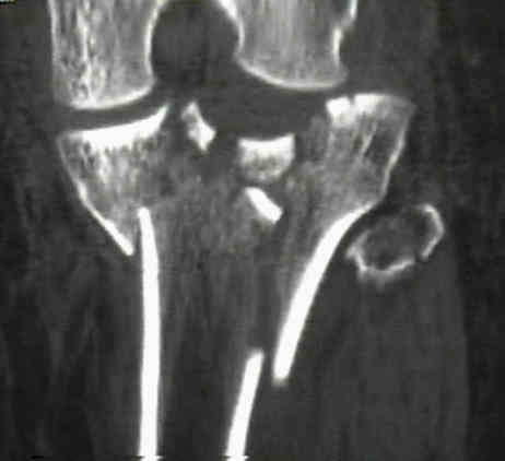
- consider CT scan to clearly define fracture patterns;
- soft tissue evaluation:
- pay attention to abrasions, bruising, and hemarthrosis since these are risk factors for wound breakdown;
- w/ ORIF w/ extensive periosteal stripping may result in a 20% incidence of wound breakdown & infection (some small series
report even higher rate of infection) that often leads to poor clinical results;
- compartment syndrome:
- insist on general anesthesia inorder to avoid dips in blood pressure (which occurs with spinal anesthesia) and inorder to allow
for immediate neurovascular exams;
- reference:
- Influence of Prior Fasciotomy on Infection After Open Reduction and Internal Fixation of Tibial Plateau Fractures.
- Timing of internal fixation and effect on Schatzker IV-VI tibial plateau fractures.
- Timing of definitive fixation of severe tibial plateau frx with compartment syndrome does not have an effect on rate of infection.
- Spanning fixators and surgical timing:
- Staged management of high-energy proximal tibia fractures (OTA types 41): the results of a prospective, standardized protocol.
- Compartment syndrome in Schatzker type VI plateau fractures and medial condylar fracture-dislocations treated with temporary external fixation..
- Timing of internal fixation and effect on Schatzker IV-VI tibial plateau fractures.
- Infection after spanning external fixation for high-energy tibial plateau fractures: is pin site-plate overlap a problem?
- The effect of knee-spanning external fixation on compartment pressures in the leg.
- External Fixation and Temporary Stabilization of Femoral and Tibial Trauma
- Staged Management of High-Energy Proximal Tibia Fractures
- Operative Technique
- indirect reduction stratedgy:
- consider using universal femoral distractor for assistance of reduction thru ligamentotaxis;
- condylar reduction can be improved w/ percutaneously applied reduction forceps;
- external fixation: (see external fixation for tibia fractures / circular wire fixators)
- CT scan is important for preoperative planning;
- femoral distractor should be considered;
- fracture reduction: need reduction before fixator placement;
- percutaneous incision allows for insertion of periosteal elevator to elevate depressed fragments;
- fracture clamps allows percutaneous fracture stabilization;
- pin placement: (see safe zone)
- transfixing olive wires are placed in the proximal fragments with use of orientation of main fracture lines as seen on the
preop CT scan;
- 1st wire: inserted through fibular head from lateral to medial
- 2nd wire: olive wire is inserted from posteromedial to anterolateral;
- pitfalls:
- prevent the leg from "sagging" posteriorly and coming into contact with the rings by padding the rings posteriorly;
- proximal wires need to be 1 cm from the joint line to avoid transgressing the reflections of the joint capsule
- references:
- Open reduction and internal fixation compared with circular fixator application for bicondylar tibial plateau fractures.
- Open reduction and internal fixation compared with circular fixator application for bicondylar tibial plateau fractures. Surgical technique.
- Indirect reduction and hybrid external fixation in management of comminuted tibial plateau fractures
- External Fixation and Limited Internal Fixation for Complex Fractures of the Tibial Plateau.
- Internal versus External Fixation of Bicondylar Tibial Plateau Fractures.
- Treatment of bicondylar tibia plateau fractures using locked plating versus external fixation
- open reduction stratedgy:
- single anterior incision (which is compatible with a TKR incision for the future) vs lateral and posteromedial incisions (better
for wound healing);
- references:
- Complications associated with internal fixation of high-energy bicondylar tibial plateau fractures utilizing a two-incision technique.
- Treatment of complicated tibial plateau fractures with dual plating via a 2-incision technique.
- The use of an anterior incision of the meniscus for exposure of tibial plateau fractures requiring open reduction and internal fixation.
- Anterior Approach to the Knee with Osteotomy of the Tibial Tubercle for Bicondylar Tibial Fractures.
- Combined Anterior and Posterior Approaches for Complex Tibial Plateau Fractures.
- Early wound complications after operative treatment of high energy tibial plateau fractures through two incisions.
- Treatment of bicondylar tibia plateau fractures using locked plating versus external fixation
-
- consider performing complete fasciotomy;
- fixation stratedgy:
- k wire fixation:
- k wires are inserted to maintain provisional fixation;
- take care that k wire position does not interfere with plate application;
- ultimate goal is to have a synthese lateral locking plate with medial washer to provide fixation for both plateau frx;
- medial plateau:
- usually fixation of the medial plateau is easier than the lateral plateau;
- consider temporary fixation of the medial w/ a simple medial butress plate;
- even if there is a coronal split into the medial plateau, the butress plate will allow a near anatomic reduction which
then allows fixation of the lateral plateau using the medial joint line as a reference;
- posteromedial incision (for secondary coronal plane fracture);
- plane between the semitendinosis and gastrocnemius
- ref: Posterior coronal plating of bicondylar tibial plateau fractures through posteromedial and anterolateral approaches in a healthy floating supine position.
- references:
- Frequency and fracture morphology of the posteromedial fragment in bicondylar tibial plateau fracture patterns.
- Stabilization of the posteromedial fragment in bicondylar tibial plateau fractures: a mechanical comparison of locking and nonlocking single and dual plating methods.
- Posteromedial second incision to reduce and stabilize a displaced posterior fragment that can occur in Schatzker Type V bicondylar tibial plateau fractures.
- lateral plateau: (see synthes plates)
- lateral locking plate is applied in the usual manner;
- once the lateral plate proximal anterior and posterior locking screws are applied, the medial buttress plate is removed,
allowing a medial washer to be inserted over the central proximal screw;
- be cafeful of use of isolated lateral locking plate with posteromedial frx with a predominantly coronal fracture line;
- references:
- Fracture pattern and fixation type related to loss of reduction in bicondylar tibial plateau fractures.
- Single lateral locked screw plating of bicondylar tibial plateau fractures.
- wound closure:
- expect that anterior compartment swelling will interefere with wound closure;
- consider proximal wound closure and leaving the distal half of the wound open to prevent compartment syndrome;
- "pie crust" technique is a simple technique to facilitate delayed wound closure;
- consider wound vac +/- bead pouch;
- ref: Multiple relaxing skin incisions in orthopaedic lower extremity trauma.
- IM Nailing:
- Retropatellar nailing and condylar bolts for complex fractures of the tibial plateau: Technique, pilot study and rationale.
- A comparative study for complex tibial plateau fractures: nailing and compression bolts versus modern and traditional plating.
- Biomechanical comparison of intramedullar versus extramedullar stabilization of intra-articular tibial plateau fractures.
- Patella osteotomy: a new approach for complex trauma around the knee.
- Post Operative Care and Compications:
- Complications of High-Energy Bicondylar Tibial Plateau Fractures Treated with Dual Plating Through Two Incisions.
- varus deformity is common w/ type V frx;
- references:
- Complications after tibia plateau fracture surgery.
- [Nicotine in plastic surgery: a review]
- Recovery of knee function following fracture of the tibial plateau.
- vascular complications:
- Evaluation of Popliteal Artery Injury Risk With Locked Lateral Plating of the Tibial Plateau
- Injury to the Anterior Tibial System During Percutaneous Plating of a Proximal Tibial Fracture
Staged management of high-energy proximal tibia fractures (OTA types 41): the results of a prospective, standardized protocol.
Treatment of Complicated Tibial Plateau Fractures With Dual Plating Via a 2-incision Technique
Patella Osteotomy: a new approach for complex trauma around the knee
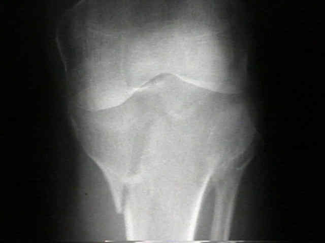
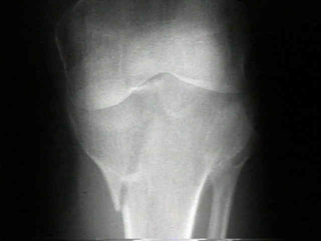
- case example:
- 40-year-old female involved in MVA, sustaining bicondylar tibial plateau frx, but no other injuries;
- interesting points about this case:
1) the initial AP of the knee did not adequately show the lateral plateau frx, since the knee immobilizer had been left in place;
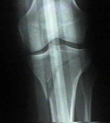

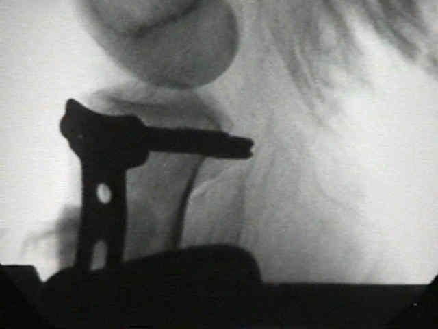
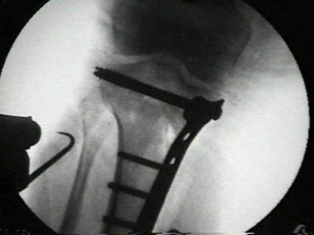
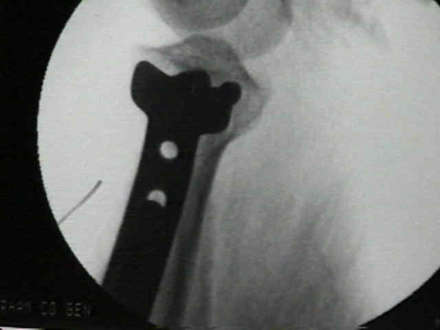
2) because the medial plateau was more comminuted and displaced than the lateral plateau, the surgeon decided to apply a "T" butress plate to the medial side w/ two proximal 6.5 mm cannulated screws angled slightly posteriorly to engage the lateral plateau frx;
- case example:
