
- See:

- Enhancement of Fixator Stability
- Ilizarov Menu
- Ring characteristics
- Safe Zone of Pin Insertion
- Foot Inclusion
- Synthes Hybrid Fixator
- Wire Insertion Technique
- Discussion:
- in the report by Paley D and Maar DC (2000), the authors report on 19 patients treated with Ilizarov devices;
- mean bone defect was 10 cm in length, and the mean external fixation time was 16 months;
- 10 patients required debridement of the bone ends and/or bone grafting;
- functional results were graded as 12 excellent, 6 good, and one poor;
- there were 22 minor complications and 16 major complications;
- Ilizarov bone transport treatment for tibial defects.
- PreOp:
- Exam:
- knee ligament injuries:
- skin abrasions / open fracture
- distal sensation and pulses (check dp while compressing pt and vice versa);
- consider compartment syndrome
- ankle range of motion;
- Radiographs:
- length of space between tibial plateau and fracture;
- length of fracture;
- length of space between fracture and tibial plafond;
- associated fibular fractures;
- Templating: 
- use opposite normal extemity to help template ring sizes;
- two finger breadths of clearances are required;
- more space is required posteriorly than anteriorly;
- space between inner rings must be > length of fracture comminution
- assess potential for transfibular wires (contra w/ fractures)
- w/ open or distal frx consider need for foot inclusion;
- Frame Construction:
- in most cases, the frame should be inserted preoperatively to save time;
- two half rings are selected that are 2-3 cm larger than the major diameter of the injured limb (less space is needed anterior to the tibia);
- the rings are positioned in the same plane and a bolt and nut anchor together both ends of the half rings;
- typically, a 4 ring assembly in required w/ 2 rings proximal and distal to frx site;
- most proximal ring in tibial mounting can be open section ring attached to complete ring, allowing maximum flexion & providing two levels of fixation;
- two rings are used on large frags, & ring & drop post used for smaller fragments;
- two proximal & distal rings are connected via two telescoping rods of appropriate length (the inner rings must span frx comminution);
- threaded rods:
- initially, only a single long anterior and posterior threaded rod is used to connect the rings together;
- this leaves plenty of space medially and laterally for insertion of wires and half pins;
- once fixation is complete, two additional threaded rods can be placed;
- plane for fracture site compression:
- the configuration should be planned in such a way that compression can be applised between the ring which span the fracture site;
- consider using the special quadrangular nuts to connect the threaded rods;
- these nuts will help control even compression across the frx site;
- Frame Construction - Proximal Fractures:
- a 5/8 ring can be mounted on top of a complete ring, w/ three points of fixation using threaded hexagonal sockets;
- the 5/8 ring can be positioned just below the joint line (or at the level of the fibular head), but will still allow knee flexion;
- alternatively, two 5/8 rings can be stacked together (connected by hexagonal sockets) and these are then connected to the distal rings w/ arch connetors;
- the disadvantage of this configuration, however, is that the 5/8 ring has less stability than a full ring (and therefore a 5/8 ring should generally be attached to a full ring for optimal stability);
- in the report by Geller J, et al, the authors stress the importance of anterior
placement of the oblique tension wires in the proximal tibia inorder to resist the forces occuring in the saggital plane;
- the angle of intersection of these wires should be greater than 60 deg for optimal stability;
- Tension wire position for hybrid external fixation of the proximal tibia.
- Frame Construction - Distal Fractures:
- w/ minimal plafond displacement, 3 sets of wires & rings are used:
- one just above the plafond, the other in proximal tibia, and third through the os calcis.
- some distraction is possible between 2 distal rings, and reduction of metaphyseal fragments is facilitated
by application of tension to wires with stop nuts;
- Surgical Strategy:
- preliminary reduction of fracture;
- insertion of transverse proximal and distal wires wires perpendicular to the knee and ankle joint lines;
- application of frame;
- tensioning of proximal and distal wires;
- frame brought to the wires (wires are not brought to the frame);
- this achieves partial reduction in the coronal plane and helps to suspend the leg in the middle of the frames;
- application of distraction across the fracture site if shortening is present;
- Surgical Technique:
- positioning:
- supine position, hip bump, and flouro on opposite side of table;
- the combination of a large hip bump and a sterile "foot bump" will create a large berth underneath the leg which facilitates insertion of wires and application of the frame;
- reduction:
- traction will usually achieve an approximate reduction;
- use Russe method to ensure proper rotation (tubercle to bi-malleolar axis);
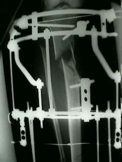
- application of frame:
- open the frame on one side (like a book) and place around the leg;
- coupling bolts are aligned parallel to the crest of tibia;
- ensure that there is proper clearance with at least one fingerbreadth of space anteriorly and two finger breadth of clearance posteriorly;
- too much clearance, however, dramatically reduces the stiffness of the construct;
- the leg can be suspended within the rings by using suction tubing tied across the bottom of the leg and over the top of the ring;
- coronal wires and frame attachement:
- see: safe zone of pin insertion and wires insertion techniques:
- proximal coronal plane reference wire:
- wire is placed at level of & parallel to knee joint and marked;
- a wire is then placed approx one cm below joint line and marked;
- distal coronal plane wire:
- is placed prior to the remaining proximal wires;
- a wire is placed transverse to ankle joint and marked;
- the distal wire is driven across the fracture site;
- frame attachment: frame is attached to the proximal and distal wires;
- mid-shaft wires:
- w/ residual displacement at the frx site, olive wires can be inserted on opposite sides of the frx and are tensioned until frx reduction is achieved; 
- remaining proximal wires:
- medial face wire:
- inserted from posteromedial side of tibia to antero-lateral side;
- flexing the knee may help avoid the pes anserinus;
- transfibular
- is driven across tibia to exit on anteromedial surface;
- ensure that this wire is not too distal so as to have the drop post encroach on the fracture site;
- remaining distal wires:
- see: safe zones and wires insertion techniques:
- ensure that there is proper rotational alignment;
- ensure that rings remain centralized;
- ensure that the fracture is reduced;
- use the Russe method to measure the bi-malleolar axis (measured off the tubercle) on the normal leg to help judge rotation off the fractured leg;
- remaining wires: are attached to remaining rings;
- half pins: half pins are attached to appropriate rings;
- Post Op:
- calcaneal wires are removed at six weeks, & ROM exercises are started;
- in cases w/ severe articular comminution, proceed w/ second stage at about 15 days when the soft tissues were healed

Limb Reconstruction by Free-Tissue Transfer Combined With the Ilizarov Method.