
- See: extenal rotators:
- Discussion:
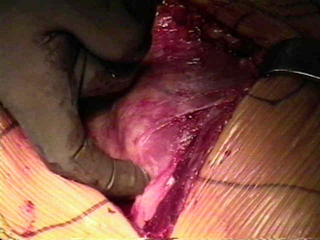
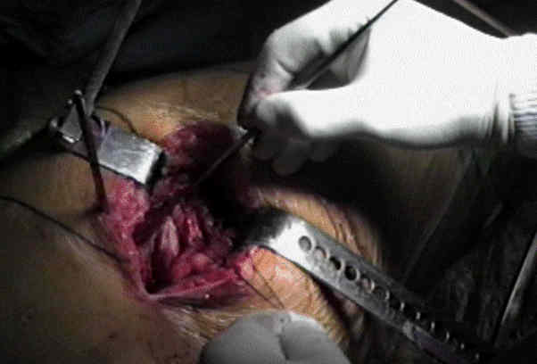
- divide trochanteric bursa & bluntly sweep it posteriorly to expose short external rotators & posterior edge of medius;
- anatomy:
- from superior to inferior external rotators include:
- piriformis, gemellus superior, obturator internus, gemellus inferior, and quadratus femoris;
- note posterior border of medius is almost in line w/ femoral shaft and anterior border fans anteriorly;
- positioning:
- flex knee & internally rotate hip to place short external rotators under tension;
- this also pulls the operative field farther from the sciatic nerve;
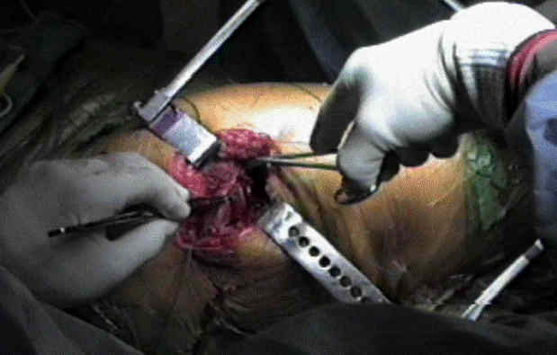
- place a blunt homan retractor underneath the medius (and over the anterior acetabular labrum) and retract the medius anteriorly;
- this will help expose the superior capsule;
- palpate sciatic nerve as it passes superficial to obturator internus and the gemelli;
- sciatic nerve emerges below piriformis & passes downwards superficial to obturator internus & gemelli & quadratus femoris;
- palpate tendinous insertions of piriformis and obturator internus and place tagging suture in the tendon for identification at closure;
- tag piriformis tendon along w/ conjoined external rotators;
- piriformis tendon, obturator internus tendon (conjoint w/ gemelli tendons) & tendon of obturator externus are identified & freed
w/ electrocautery from their insertions at greater trochanter;
- detach muscles close to femoral insertion & reflect backward laying them over sciatic nerve to protect it during the case;
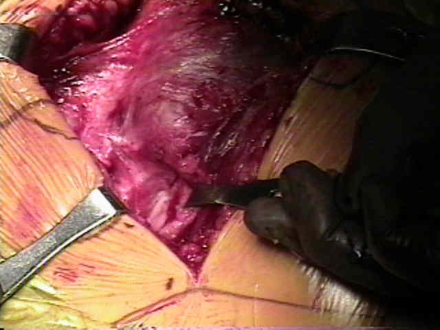
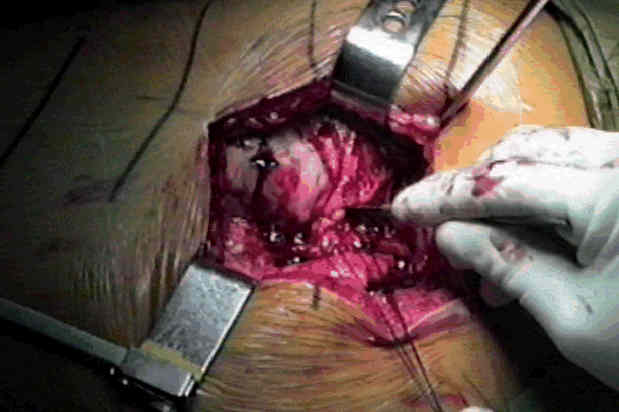
- divide external rotators, including at least proximal half of quadratus femoris, at their insertion on femur;
- branches of MFCA emerge above & below quadratus femoris;
- keep a retractor on cut edge of external rotators for protection;
- keep folded posteriorly to protect the sciatic nerve;
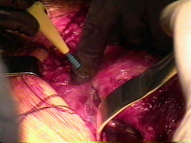
- develop plane between these muscles & underlying capsule;
- branches of MFCA emerge above & below quadratus femoris to anastomose w/ inferior gluteal vessels & ascending branches from
profunda femoris (cruciate anastomosis) and can be ligated as required;
- quadratus femoris is incised close to quadrate tubercle exposing terminal branch of medial femoral circumflex artery, which lies deep
to proximal third of quadratus femoris, accompanying inferior border of the obturator externus;
- reflect short external rotators posteriorly protecting sciatic nerve;
- turning medial cut ends of muscles backwards over nerve helps to protect it