Ortho Preferred Malpractice Insurance
Home » Joints » Shoulder » Arthroplasty of the Shoulder

Arthroplasty of the Shoulder

- Discussion:

- Total Shoulder Arthroplasty:    (considerations: hemiarthroplasty vs TSA)
- Reverse arthroplasty

- Indications:
- indicated for end stage DJD or RA shoulder involvement;
- contra-indications to shoulder arthroplasty include combined rotator cuff / deltoid paralysis
and recent joint infection;
- in these rare circumstances, arthrodesis may be considered;
- may occur as a consequence of recurrent shoulder instability;
- infection control
- references:
- Shoulder Arthroplasty in Patients with a Prior Anterior Shoulder Dislocation. Results of a Multicenter Study.
- Dislocation arthropathy of the shoulder 

Anatomical total shoulder replacements are in widespread use for the treatment of patients with arthritic shoulders. Over many years the designs have changed in light of outcomes and research, particularly related to failure mechanisms. An intact rotator cuff insertion is now recognised as essential for optimal outcome from an anatomical configuration shoulder replacement. This is because one of the functions of the rotator cuff is to depress the humeral head and centre it in the glenoid during shoulder movement, particularly elevation. In the absence the superior rotator cuff tendon supraspinatus, the humeral head rides up and migrates proximally such that it may articulate with the under surface of the acromion. This will mean there is point loading on the superior edge of a prosthetic glenoid component resulting in excessive eccentric wear and the ‘rocking horse effect’ resulting in loosening of the component. While analgesia may be adequate, function is likely to be poor and risk of glenoid component wear and loosening and hence failure is high.

Many Orthopaedic companies have developed such shoulder replacement implants.  In this case, I am using the stemless humeral implant on the anatomic system from the Italian company Lima. The stemless humeral component is constructed on a 3D printer to create a single piece of trabecular titanium. Hip surgeons have used the same trabecular titanium for successful reconstruction of the hip acetabulum with impressive bony ingrowth of the implant. In the shoulder, if the patient’s proximal humeral metaphyseal bone is not too soft then impaction of the trabacular titanium core humeral implant gives excellent primary fixation and promotes future bone ingrowth securing the platform component.

This is a bone preserving procedure avoiding the need to use an intramedullary stem. The same humeral core component is used for reverse total shoulder replacement as also described in Orthoracle and this facilitates revision of an anatomic replacement to reverse. The original Lima anatomic shoulder replacement uses a metalback glenoid component, fixed with press-fit and cancellous screws, into which the polyethylene component is inserted. It is very straightforward to perform revision surgery to switch the articulating components leaving the glenoid baseplate and humeral core implants, which should be solidly fixed to bone, in place.

There have been concerns with metal glenoid baseplates concerning eccentric wear of the polyethylene resulting in the humeral head component articulating with the metal rim of the baseplate. Dissociation of the polyethylene liner from the metal baseplate has also been recognised. To reduce the risk of such problems Lima have developed the TT hybrid glenoid component. The majority of the implant is polyethylene with a central peg attached to a trabecular titanium (TT) core and two much smaller peripheral pegs to ensure rotational stability which can be fixed with a tiny volume of cement, hence the hybrid label. The central TT peg is impacted into the prepared glenoid and will encourage bone ingrowth just as the humeral core implant.

If in the future revision surgery from anatomic to reverse is required, then the system has been designed to allow the polyethylene to be drilled and dissociated from the central TT core and discarded. The central peg core is firmly embedded in the glenoid into which a male to female metal baseplate can be inserted onto which the glenosphere is fixed for the reverse geometry shoulder replacement. 

Readers will also find of interest the following related OrthOracle operative techniques:

Lima stemless reverse shoulder replacement

Lima SMR Reverse Total Shoulder Replacement for proximal humeral fracture

Lima stemmed SMR reverse geometry shoulder replacement (nickel free implants)

Proximal humeral replacement: Mutars reverse geometry shoulder (Implantcast).

Clinicians should seek clarification on whether any implant demonstrated is licensed for use in their own country.

In the USA contact: https://www.fda.gov/medical-devices/products-and-medical-procedures

In the UK contact:  https://www.gov.uk/government/organisations/medicines-and-healthcare-products-regulatory-agency

In the EU contact: https://www.ema.europa.eu/en/human-regulatory/overview/medical-devices 

Read more »

This overview is brought to you by Orthoracle - the online e-learning Orthopeadic Surgery Atlas

Take the Tour

View this procedure on OrthOracle.com | OrthOracle is a commercial partner of Data trace LLC


- Exam:

- note the degree both preoperatively and postoperatively of scapulothoracic to glenohumeral motion;
- as noted by Friedman RJ (1997), patients with DJD of the shoulder reverse the normal 1:2 ratio of
scapulothoracic to glenohumeral motion ratio (this is not changed w/ arthroplasty);
- excessive external rotation:
- may indicated deficiency of subscapularis
- restricted external rotation:
- may indicated severe wear of the posterior glenoid, in which case the glenoid may have to be
reamed to a more neutral version;
- reference
- Prospective analysis of total shoulder arthroplasty biomechanics.


- Radiographs:
- AP radiograph in internal and external rotation
- axillary view to assess glenoid deficiencies;
- even if glenoid appears normal on the axillary view, any posterior subluxation may indicate excessive
poserior glenoid wear;
- CT scan:
- may allow better assesment of the glenoid version and possible posterior glenoid erosion;
- ref: The use of computerized tomography in the measurement of glenoid version.
- osteoarthritic changes:
- prominent osteophyte at the inferior margin of the humeral head or glenoid is characteristic;
- mild arthrosis: inferior osteophyte less than 3 mm in length;
- moderate arthrosis: inferior osteophyte between 3-5 mm in length, irregularity of the joint line and subchondral sclerosis;
- severe arthrosis: inferior osteophyte measuring more than 5 mm or if there is joint incongruity;
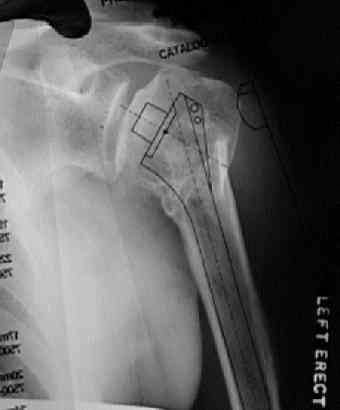
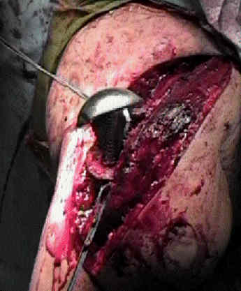

- Surgical Technique:
- anterior approach to shoulder
- hemiarthroplasty  - preparation and insertion of hemiarthroplasty
- total shoulder arthroplasty:
- glenoid component:
- total shoulder arthroplasty - video
- technical considerations with fracture
- reverse arthroplasty


- Post Operative Rehab:
      - immediate begin an active assisted range-of-motion program emphasizing forward elevation and external rotation to the side;
- active strengthening should not begin for 6 weeks postoperatively to allow the subscapularis tendon repair time to heal;
- reference:
- Rehabilitation after total shoulder arthroplasty.
- Is a formal physical therapy program necessary after total shoulder arthroplasty for osteoarthritis?

- Complications of Shoulder Arthroplasty