
- Discussion:
- see: elbow dislocations in children:
- elbow dislocation is the second most common major joint dislocation;
- dislocation is usually closed and posterior;
- mechanism:
- dislocations of elbow usually result from fall onto extended elbow.
- anatomic morphology of semilunar notch may predispose to elbow dislocation;
- central angle of semilunar notch is significantly larger in group of pts who had dislocation of the elbow compared to normals;
- classification:
- elbow dislocations without fracture are termed "simple." whereas dislocations with frx are termed complex;
- dislocations are classified according to direction of dislocation: posterior, posterolateral, posteromedial, lateral, medial,
or divergent;
- ref: Posteromedial Elbow Dislocations w/o Osseous Lesions. Characteristics, Tissue Injury Patterns, Treatments, and Outcomes
- simple dislocation: pathoanatomy:
- rupture of capsule, rupture of MCL, lateral ligaments, rupture of flexor pronator mass and less commonly, injury to
brachialis muscle;
- LCL may be the essential lesion in recurrent or persistent instability following simple dislocations of the elbow
- rupture of brachial artery has been reported;
- complex dislocation:
- dislocation w/ radial head frx: most common complex dislocation;
- frx dislocation w/ MCL injury: radial head frx & MCL Instability:
- terrible Triad: (dislocation, cornoid process frx, and radial head frx)
- ref: Classification and evaluation of recurrent instability of the elbow.
- stability of elbow:
- primary stabilizers
- MCL is the main stabilizer of the elbow joint (provides 54% valgus stability, while osseous articulation provides 33%);
- ulnohumeral articulation
- coronoid: 50% intact coronoid requirement for stability with or w/o ligamentous integrity
- olecranon contribution to stability inversely correlated with resection amount: >30% articular surface of olecranon
needed for stability
- secondary stabilizers
- radiohumeral articulation (most important)
- capsule: greatest role in extension of elbow, insignificant role (<10%) in flexion
- musculature (dynamic)
- ref: Articular and ligamentous contributions to the stability of the elbow joint.
- Exam:
- vascular injury:
- closed dislocations are rarely assoc w/ vascular injury, whereas open &/or ant dislocations are commonly assoc w/ such injury;
- in open dislocations, brachial artery is disrupted by forcible hyperextension (median nerve injury is commonly associated with
such injuries);
- reference:
- Closed elbow dislocation and brachial artery damage.
- Occult closed posterior elbow dislocation with intimal rupture of the brachial artery in a 71-year-old male
- neuro injury:
- diff dx: compartment syndrome: before assuming that a nerve injury is present consider whether there is an evolving
compartment syndrome;
- remember that the pain of the compartment syndrome is distracted by the more obvious elbow dislocation;
- these patients often have had conscious sedation, which can blunt on going pain from a compartment syndrome;
- after several hours, the acute pain of a compartment syndrome may diminish (nerve ischemia), after which it will be
difficult to distinguish from a nerve injury;
- neuropraxia is occurs in 20%, usually involving ulnar or median n (AIN branch);
- ulnar nerve palsy may occur up to 14% of adult elbow dislocations, and the occurance of ulnar nerve palsy is much higher in
pediatric dislocations w/ an associated medial epicondyle frx;
- most neurologic deficits are transient, but entrapment of median nerve w/ elbow joint after manipulation is more common in
pediatric dislocations;
- reference:
- Median nerve palsy after posterolateral elbow dislocation.
- bony displacement:
- when nl elbow is extended, olecranon process & medial & lat form 3 points on straight line, & when nl elbow is flexed to 90 deg
in lateral view, olecranon is aligned vertically w/ epicondyles;
- tip of the olecranon is, however, definitely posterior to the plane of the epicondyles;
- in post dislocation, olecranon process is displaced backward from its normal position in relation to humerus, & one can palpate
the concavity of the semilunar notch;
- increasing degree of elbow flexion exaggerates the prominence of the olecranon process
- very important to examine whole upper extremity for evaluation of essex-lopresti lesion at wrist or associated fractures
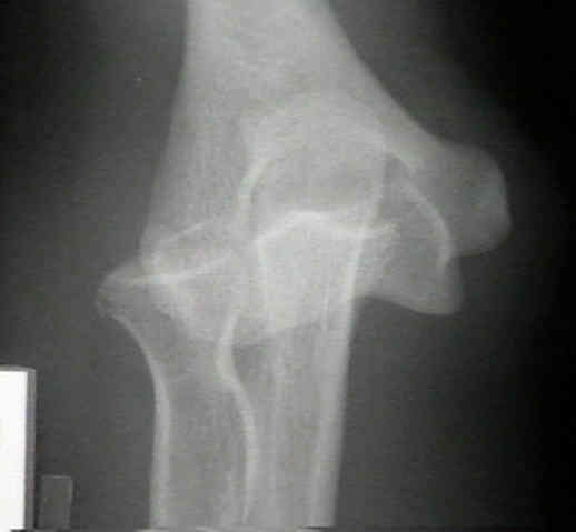
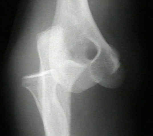
- Radiographs:
- it is essential to obtain radiographs both before and after reduction in order to asses for associated fractures (indicating
a complex dislocations);
- note the radial head avulsion frx, seen in this example

- Reduction of the Posterior Dislocation:
- Post Reduction Radiographs and Assessment of Stability:
- generally the elbow will be stable in 90 deg or more of flexion;
- the question is whether the elbow will be stable upto 30 deg flexion;
- if instability occurs in 30 deg of flexion, then place forearm in maximum pronation which maximizes the stress on the
MCL which reduces posterolateral subluxation;
- if there is increased stability in pronation, then the elbow should be placed in a cast brace with the elbow in pronation;
- after clinically determining that the reduction will not be lost in 30 deg of flexion, obtain a portable lateral and AP radiograph;
- look for joint widening, joint irregularity, or malalignment;
- in difficult cases, flouroscopy can be used;
- in cases of simple dislocation, persistent instability as the elbow is extended may indicate interposition of soft tissue or an
osteochondral fragment;
- Non Operative Treatment:
- stable articulation will allow for early flexion & extension if valgus stress is prevented after reduction;
- no one has demonstrated a benefit from operative repair of MCL in simple dislocations;
- best Rx results are obtained w/ early protected ROM begun before 2 wks;
- if there is increased stability in pronation, then the elbow should be placed in a cast brace with the elbow in pronation;
- final clinical outcome for simple dislocations of the elbow is dramatically affected by the duration of immobilization;
- recurrent dislocation is unusual;
- mild loss of extension is common, prolonged immobilization over two wks is assoc w/ greater flexion contracture;
- reference: Simple elbow dislocation among adults: a comparative study of two different methods of treatment.
- Operative Treatment of Simple Dislocations:
- redislocation of elbow w/ passive range of motion or redislocation in plaster implies severe valgus instability w/ rupture of both
MCL & flexor forearm muscles;
- under these circumstances, operative treatment is indicated;
- repair of MCL may be attempted but is not guaranteed to restore stability;
- consider use of a hinged elbow fixator, which will allow early range of motion as well as stability;
- arthroscopy: when exam findings unconvincing, possibility of intra-articular lesion, and reveals radiohumeral joint laxity
- Management of Complex Elbow Dislocations:
- dislocation w/ radial head frx
- terrible triad
- Complications:
- valgus instability:
- patients will show a variable amount of MCL laxity which correlates with a worse clinical and radiographic result;
- to maximize the stress on the medial collateral ligament, the forearm should be placed in full pronation, which
reduces the posterolateral subluxation;
- posterolateral instability;
- heterotopic ossfication
- whether or not all patients with simple elbow frx dislocations should receive prophylaxis is a matter of controversy;
- chronic dislocations:
- in some cases, recurrent instability will be due to posterolateral instability;
- management of untreated posterior dislocations of the elbow three or more wks after injury may require open reduction;
- posterior approach: w/ lengthening of triceps, removal of fibrous tissue, & possible K-wire stabilization has been
recommended.
- in the report by H. Moritomo et al. 1998, the authors discuss reconstruction of the coronoid process (w/ graft taken from the
olecranon) in order to help block dislocation;
- Reconstruction of coronoid process for chronic dislocation of the elbow. Use of a graft from the olecranon in two cases.
Surgical vs non-surgical treatment of ligamentous injuries following dislocation of the elbow joint. A prospective randomized study.
Simple dislocation of the elbow in the adult. Results after closed treatment.
Ligamentous injuries in dislocations of the elbow joint.
Posterior dislocation of the elbow.
Posterior dislocation of the elbow in children.
Fractures and dislocations about the elbow in the head-injured adult.
Elbow dislocation in children and adults. A long-term follow-up of conservatively treated patients.
Posterior dislocation of the elbow.
Recurrent dislocation of the elbow.
Dislocations of the elbow and intraarticular fractures.
Elbow subluxation and dislocation. A spectrum of instability.