
- See:
- Flexion and Extension Views:
- Technique:
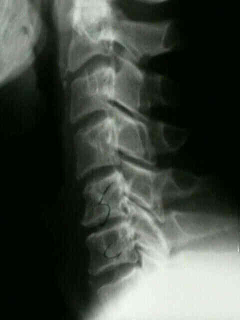
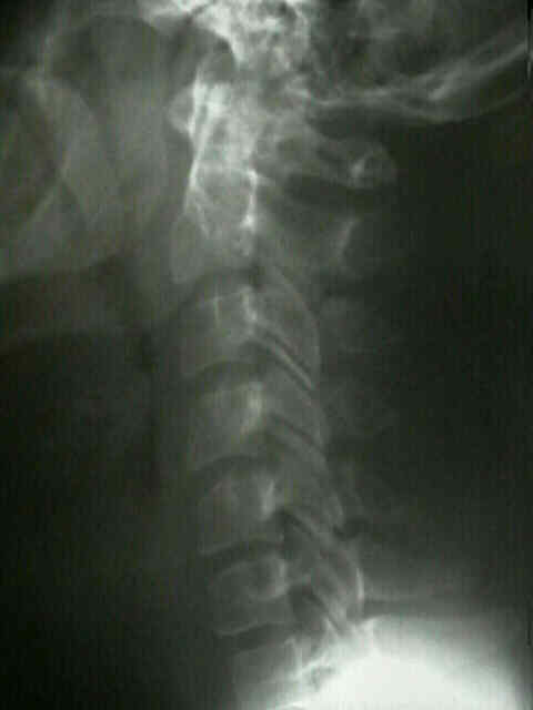

- Upper Cervical Spine:
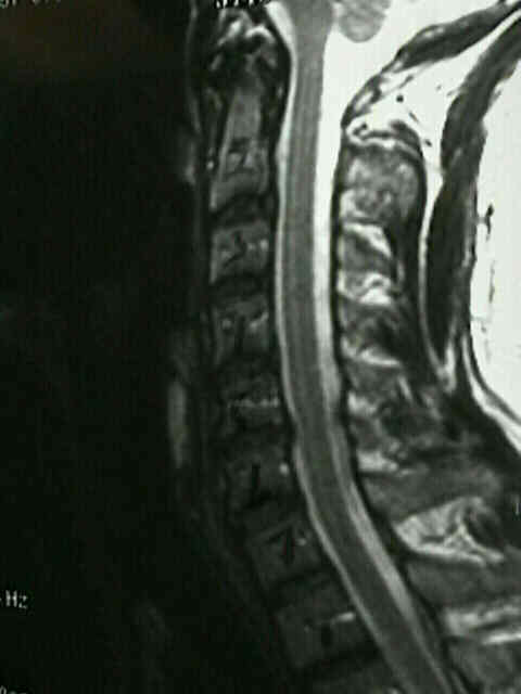
- prevertebral soft tissues
- occipital-atlanto-axial injury:
- atlanto-occipital disassociation
- C1-C2 interspinous space should not be greater than 10 mm;
- atlanto-axial impaction (rheumatoid C-spine)
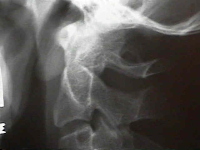
- atlas (Jefferson frx)
- axis (odontoid frx / hangman frx)
- atlantoaxial distance & SAC;
- ADI in children (< 10 yrs) < 3.5 mm; (see pediatric C-spine)
- ADI in adults < 3 mm;
- an anterior shift of C1 on C2 of more than 3-5 mm implies injury to transverse ligament (see atlanto-axial subluxation);
- shift > 5 mm implies injury transverse & alar ligaments;
- SAC:
- greater than 18 mm is normal normal;
- 15-17 mm - grey zone;
- less than 14 mm is consisent w/ cord compression;
- pseudosubluxation of c spine:
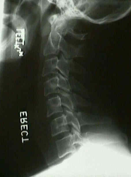
- Sub-Axial Spine - Alignment:
- posterior cortices: (more important than anterior cortices)
- anterior or posterior translation of vertebral bodies > 3.5 mm implies instability; 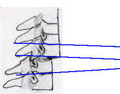
- w/ less than 25% relative shift of one vertebral body over another consider facet frx;
- w/ 25% relative shift consider unilateral facet dislocation and w/ 50% shift, consider or bilateral facet dislocation;
- vertebral body angulation / translation:
- patterns of instability include:
- 1.7 mm or greater of disk widening;
- 3.5 mm of translational displacement;
- angulation between two adjacent vertebra of 11 deg more than contiguous cervical vertebrae;
- measurements are made from each inferior endplate;
- anterior cortices:
- anterior subluxation
- minimal compression frx of anterior vertebral bodies;
- tear drop sign: bone chip off antero-inferior aspect;
- may indicate displacement of disc or posterior fragment of vertebral body into spinal canal & cord injury;
- spinolaminar line (dorsum of lateral masses) (see oblique view);
- facet joint widening;
- rotation of the facets on lateral view;
- parallel articular process facets;
- spinous process angulation:
- C1-C2 interspinous space should not be greater than 10 mm;
- widening is present when the distance is more than 1.5 times the inter-spinous distance of adjacent spinal segments;
- fanning implies middle column disrupton
Biomechanical analysis of clinical stability in the cervical spine.
Neurapraxia of the cervical spinal cord with transient quadriplegia.