
- See: Extensor Tendon Lacerations:
- the extensor apparatus is a "plexus of tendons with an aponeurotic sheet." - from Stack, 1962.
MP Joint Level: 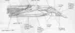

- extensor digitorum communis: (at the MP joint)
- there may be some adhesion to the joint capsule, but there is no formal insertion;
- the deep portion of the extensor tendon becomes adherent to and supplements the dorsal roof of the MP joint capsule;
- these fibers may help centralize the extensor tendons, but probably do not contribute to finger extension;
- other argue that this insertion is of functional significance;
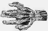
- The Anatomy of the Extensor Apparatus of the Fingers
Tubiana R, and Valentin P. Surg Clin North Am. 1964;44:897.
- at the MCP joint, four tendons, one to each finger, divides into three slips;
- middle slip connects to the proximal phalanx via the lasso like saggital bands (which encircle the digit and attach to the volar plate)
- it is the only extensor of the proximal phalanx;
- middle slip goes on to attach to the dorsal surface of the 2nd phalanx;
- two lateral slips unite to attach to dorsal surface of base of distal phalanx;
- on the radial side, the lateral slip of the EDC unites w/ the lateral slip from the lumbrical to form the lateral band;
- saggital bands: (at the MP joint)
- saggital bands anchor the extensor tendon over the central MCP joint;
- saggital bands arise from volar plate and spans both sides of MP joint;
- they adhere to collateral ligaments and deep transverse intermetacarpal ligament;
- these bands function to extend the proximal phalanx;
- conjoined tendons of intrinsics and saggital band hold the extensor tendons in a central position over the MP joint;
- rupture:
- most often involves saggital hood tear of radial side of middle finger following trauma;
- patients will be unable to actively extend the digit from a flexed position (since the tendon is subluxed) but the patient will
be able to keep the digit extended after the digit is passively extended (since the tendon is relocated);
- w/ acute injury consider nonoperative treatment w/ the MP joint splinted in extension;
- interossei: (at the MP joint)
- on both sides of MCP joint, a portion of the interossei insert into the base of the proximal phalanx and joint capsule;
- the remainder of the interossei alond w/ the lumbricals insert into the extensor apparatus at the level of the proximal phalanx,
and continues distally (as the lateral bands:) to insert into the distal phalanx;
Phalangeal Level:
- divides into 3 slips;
- central slip continues on to insert into dorsum of middle phalanx;
- two lateral bands travel on either side of proximal phalanx;
- transverse fibers:
- these lie distal to the saggital bands;
- arise from the dorsal surface of the lateral bands and envelop the dorsal extensor apparatus;
- they act to flex the MP joint
- lateral bands:
Littler JW. Surg Clin North Am. 1967;47:415-432.
Muscle Function in the Fingers.
Stack HG. J Bone Joint Surg 1962;44-B:899.
The microvascular anatomy of the distal extensor tendon.
Warren RA, Kay RNM, Norris SH. J Hand Surg. 1988;13-B:161.
Common variations of the radial wrist extensors.
Albright JA. J Hand Surgery. 1978;3:134.
Variations of the extensor tendons of the fingers. Surgical significance.
SchenckRR. J Bone Joint Surg Am. 1964;46:103.