
- One Incision - Modified Henry Approach:
- see: Henry Approach to the Forearm;
- some surgeons feel that this is the prefered technique noting that modern suture anchors anchors have permitted safe repair of
biceps tendon through one anterior incision;
- advantages: direct approach, avoids PIN injury, and minimal ectopic bone formation;
- disadvantages: may injury radial nerve if surgeon attempts to pass tendon thru drill holes made in the radius;
- this is less of a problem now that stronger bone anchors are available;
- incision:
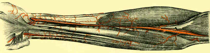
- begins either medial or lateral to biceps, extends transversely over antecubital fossa, and then extends distally over the BR;
- identify and protect the lateral antibrachial cutaneous nerve;
- anatomic variations have been described in the musculocutaneous nerve pierces the distal biceps tendon;
- muscle interval:
- exposure between the brachioradialis (w/ radial / lateral retraction) and pronator teres (medial retraction);
- limiting forceful lateral retraction of BR helps avoid injury to the PIN;
- tendon end is identified, elevated out of wound, and debrided;
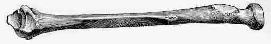
- thru the empty space (occupied by the biceps), insert a tonsil and identify the radial tuberosity (forearm fully supinated);
- saline lavage of the tendon tract to prevent heterotrophic ossification;
- in the traditional approach, deep interval lateral to the biceps tendon is developed and the leash of radial recurrent vessels is
ligated to increase the exposure;
- avoid injury to the PIN and anterior interosseous nerve;
- keep forearm supinated inorder to keep the PIN as far away as possible;
- retractors placed around the radial tuberosity are the main cause of nerve compression;
- limiting forceful lateral retraction of BR
- bone anchors:
- inserted into the ulnar aspect of the tuberosity, and reattach the tendon;
- main mistake is to position the anchor proximal to the tuberosity (needs to be in the ulnar aspect of the tuberosity);
- note the length of the bone anchor (avoid excessively long bone anchors which might penetrate the far cortex);
- references:
- The effect of drilling angle on posterior interosseous nerve safety during open and endoscopic anterior single-incision repair of the distal biceps tendon.
- The Effect of Drill Trajectory on Proximity to Posterior Interosseous Nerve During Cortical Button Distal Biceps Repair
- The pullout force for Mitek mini and micro suture anchor systems in human mandibular condyles.
- How to Avoid Posterior Interosseous Nerve Injury During Single-Incision Distal Biceps Repair Drilling
- postoperative care:
- immobilize the elbow in 90 deg flexion for 2 weeks followed by progressive increases in elbow ROM using hinged brace;
- passive pronation and supination with the elbow flexed at or greater than 90° of flexion is allowed after 2 weeks;
- active flexion is started at 8 weeks
Rupture of the distal insertion of the biceps brachii tendon.
Rupture of the distal tendon of the biceps brachii. A biomechanical study.
Rupture of the distal tendon of the biceps brachii. Operative versus non-operative treatment.
Distal biceps brachii tendon avulsion: a simplified method of operative repair.
Partial rupture of the distal biceps tendon.
Repair of the distal biceps tendon using suture anchors and an anterior approach
Single-incision repair of acute distal biceps ruptures by use of suture anchors.
Distal biceps brachii repair. Results in dominant and nondominant extremities.
A method for reinsertion of the distal biceps brachii tendon
Repair of avulsion of insertion of biceps brachii tendon.
Complications of Repair of the Distal Biceps Tendon with the Modified Two-Incision Technique.
Repair of distal biceps tendon ruptures using a suture anchor and an anterior approach.
Complications of distal biceps tendon repairs.
High complication rate following distal biceps refixation with cortical button
