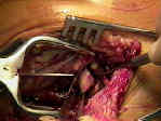
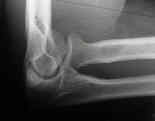
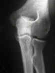
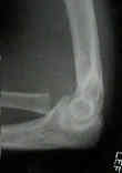
 - See:
 - Adult Radial Neck Frx / Pediatric Radial Head Frx
- Discussion:
- radial head frx is most common type of elbow fracture in adults;
- frx of the radial head occurs primarily in adults, whereas fractures of the radial neck are more
common in children;
- frx of the radial head and neck of the radius generally results from a hard fall on an outstretched hand;
- impact of fall drives head of radius axially onto capitulum of humerus;
- high frequency of frx in anterolateral aspect of radial head occurs as a result of lack of subchondral bone
under anterolaterl aspect of radial head;
- because the anterolateral aspect of radial head does not articulate w/ sigmoid fossa, frx in the region are amenable to fixation
w/ small screws;
- associated injuries:
- frx of the capitellum
- distal radius frx
- dislocation of the distal RU joint (Essex Lopresti Fracture)
- valgus instability (MCL rupture)
- probably more common than is reported;
- indications for repair of the MCL will be determined based on stability of the elbow thru a functional range of motion;
- rupture of the triceps tendon
- elbow dislocation:
- terrible triad: RHF + LCL / MCL + coronoid process frx;
- Risk of Subluxation or Dislocation after Operative Treatment of Terrible Triad Injuries.
- Terrible triad injuries of the elbow: does the coronoid always need to be fixed?
- Fixation versus replacement of radial head in terrible triad: is there a difference in elbow stability and prognosis
- Terrible Triad Injuries of the Elbow
- Outcomes of coronoid-first repair in terrible triad injuries of the elbow
- Radial head resection versus prosthetic arthroplasty in terrible triad injury: a retrospective comparative cohort study
- Radial head replacement vs reconstruction for treatment of terrible triad injury of the elbow: a review and meta-analysis
- Dx and Exam:
- dx of a radial head fracture may be difficult;
- pain, effusion in the elbow, & tenderness on palpation directly over radial head are typical manifestations;
- if frx is displaced, click or crepitus over radial head is detected w/ supination;
- if elbow ROM is limited, then aspirate and inject several cc of lidocaine, and then re-examine;
- check for blocks to flexion-extension as well as supination-pronation;
- wrist tenderness with ROM is common;
- Radiographic Features:
- AP & Lat (look for fat pad sign)
- Radiocapitellar view: forearm in neutral rotation & the x-ray tube angle 45 d. cephalad
- main disadvantage of this classification is that radiographs may underestimate the true degree of comminution;
- references:
- Magnetic resonance imaging in radial head fractures: most associated injuries are not clinically relevant.
- The cortical irregularity in transition zone of radial head and neck: a reliable radiographic sign of occult radial head fracture.
- Treatment: (based on Mason classification)
- type I
- type II
- less than 30% of radial head;
- more than 2 mm displacement
- ORIF of radial head fracture
- type III
- excision of radial head:
- radial head implants:
- references:
- Treatment of displaced segmental radial-head fractures. Long-term follow-up.
- adult radial neck fracture
- complex fractures
- radial head frx & elbow dislocation
- radial head frx & MCL instability
- Essex Lopresti Fracture
- references:
- Radial head fractures and their effect on the distal radioulnar joint. A rationale for treatment.
- Radial head fracture. A potentially complex injury.
- Radial head fractures with acute distal radioulnar dislocation. Essex-Lopresti revisited.
- Surgical Considerations:
- posterolateral approach: (Kocher Approach)
- approach the fascial plane between the ECU and anconeus muscle
- direct lateral approach is preferred by some surgeons because it spares the lateral ulnohumeral ligament;
- ORIF of radial head fracture / safe zone for implant insertion
- radial head replacement:
Fractures of the radial head treated by internal fixation: late results in 26 cases.
Internal fixation of proximal radial head fractures.
Open Reduction and Internal Fixation of Fractures of the Radial Head.


