
- See Technique of IM Nailing
- Indications:
- tibial shaft fractures within 4 cm distal to the tibial tuberosity to 4 cm proximal to the ankle may be treated with interlocking techniques;
- Soft Tissue Injury:
- open tibial frx (Gustillo classification):
- reconstruction for leg defects:
- Frx Stability:
- see Winquist classification:
- fracture is stable with at least 50% of cortical continutiy and within the middle 1/3 of the tibia
- when stability is in doubt, static interlocking allows control of alignment, length, and rotation (esp. rotation);
- Exam:
- compartment syndrome:
- note, w/ IM nailing, posterior cortex of tibia may be frxed on insertion of nail w/ possible nerve or vascular injury in deep posterior compartment;
- MCL tears are common and PCL may be injured in about 5 %;
- Templating:
- length:
- measuring length of nail is simple, a tape measures the length from medial malleolus to tibial tuberosity on unaffected side;
- nail should come w/in 2 cm of articular surface at distal end of tibia to provide for maximum fixation;
- overlie tibial nail template on the AP and lateral radiograph to help determine optimal nail entry position;
- intraoperative determination:
- use flouro to mark proximal entry position at level of tibial plateau, & distal position at physeal scar;
- pitfalls: do not measure nail length from the proximal tibial joint line but instead measure length from the nail entry site;
- use radiographic ruler on the reduced leg or on the contralateral leg;
- rotational alignment:
- use opposite leg as a reference for rotational alignment;
- Positioning for Tibial IM Nailing
- Difficult Fractures:
- proximal tibial fractures
- fractures w/ long anterior spike
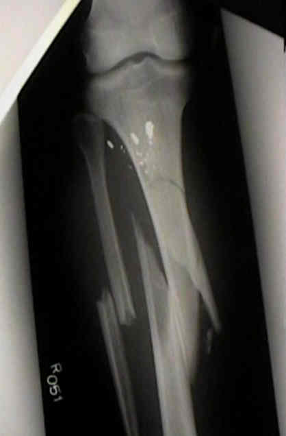
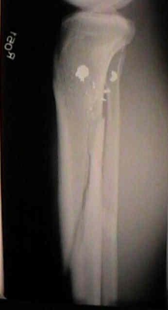
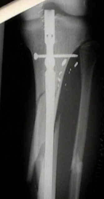
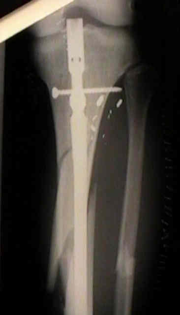
- in this case, the fracture was left malreduced (and w/ poor bony opposition) and only a single proximal interlocking screw;
- after 4 months, the proximal interlocking screw broke, the fracture shortened, and the long anterior spike caused tenting of the skin
Intramedullary nailing of fractures of the tibia in diabetics
Study finds 47% primary union rate in tibia patients with ‘critical-sized’ bone defects