
- General Discussion of IM Nails 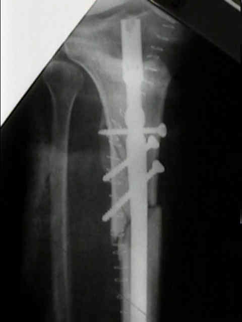
- PreOp Planning
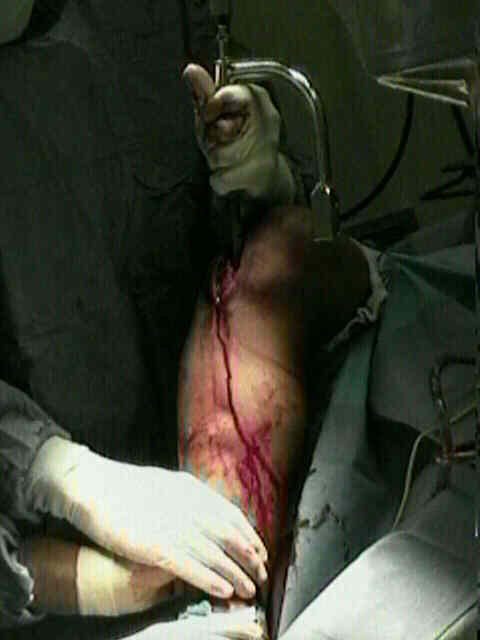
- positioning
- note rotational alignment of the opposite non injured side.
- cautions:
- proximal tibial fractures (high complication rate w/ IM nailing)
- distal tibia fracture
- IM nailing of open tibial fractures
- segmental tibia fractures: / treatment methods for tibial defects
- compartment syndrome
- w/ IM nailing, posterior cortex of tibia may be fracture on insertion of nail w/ possible nerve or vascular injury
in posterior compartment;
- narrow intramedullary canal;
- reference:
- Heat-induced segmental necrosis after reaming of one humeral and two tibial fractures with a narrow medullary canal.
- tourniquet:
- tourniquet may aid exposure but is avoided w/ reaming, as absence of blood flow increases extent of thermal necrosis;
- tourniquet may contribute to compartment syndrome and thermal necrosis;
- references:
- The use of a tourniquet when plating tibial fractures.
- Thermal necrosis after tibial reaming for intramedullary nail fixation. A report of three cases.
- anesthesic considerations:
- surgeons should insist on general anesethesia or short spinal so that the surgeon can evaluate for postop compartment syndrome;
- references:
- Does patient controlled analgesia delay the diagnosis of compartment syndrome following intramedullary nailing of the tibia?
- Differences in attitudes to analgesia in post-operative limb surgery put patients at risk of compartment syndrome.
- Compartment syndrome without pain!
- Silent compartment syndrome complicating total knee arthroplasty: continuous epidural anesthesia masked the pain.
- Patient Position and Fracture Reduction:
- before the case starts, the surgeon should have a plan to obtain frx reduction, noting the amount of help and equipement available;
- position the OR lights away from the center of the table, so that the guide wires do not touch.
- figure of four position:
- same as the arthrscopic position, with hip and knee flexed over the opposite leg;
- minimzes the angulatory displacement in the saggital plane;
- note that once the proper entry hole is established (AP view), the reduction on the lateral view is most important;
- w/ proximal or distal fractures, consider need for blocking screws;
- w/ open fractures (or cases in which a fasciotomy has been performed) a direct reduction is performed through the wound;
- direct reduction is often critical for proximal fractures;
- if the reduction is difficult to achieve, then a cannulated nail system should be used (both for reaming and for nail insertion);
- references:
- Does open reduction increase the chance of infection during intramedullary nailing of closed tibial shaft fractures?
- A positioning technique for closed intramedullar nailing of tibia fractures.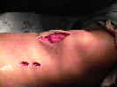
- Open Reduction and Intramedullary Nail Fixation of Closed Tibial Fractures
- Preparation for Nail Insertion: (see synthes)
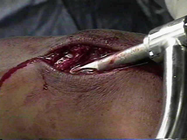
- skin incision and exposure
- Suprapatellar Intramedullary Nail Technique Lowers Rate of Malalignment of Distal Tibia Fractures.
- entry into the IM canal
- reaming of tibial fractures:
- over-reaming by 0.5 to 1 mm is probably indicated for all tibial IM nailing procedures since it helps guarantee proper nail
diameter (avoiding nail incarceration and/or posterior cortex blow out);
- tourniquet should never be inflated during reaming of the intramedullary canal, since this risks thermal necrosis (tourniquet is
rarely needed);
- avoid eccentric reaming:
- remember that the reamer follows position of guide wire and that nail follows the path left by the reamer and therefore
guide wire position needs to be carefully checked during reaming to make ensure that it is centrally located on two
radiographic views;
- eccentric reaming will cause the nail to enter into the canal eccentrically which will end up cause the distal fracture
fragment to move into varus if the reaming is eccentric laterally and will cause it to move into valgus if the
reaming is too medial;
- determine nail width:
- this is critical because under-sizing nail diameter will give a loose fit and over-sizing nail may cause nail incarceration and
resultant posterior cortex frx;
- reaming is the best way to determine nail width;
- w/ unreamed technique, sounds can be used to determine the diameter of the canal and the proper nail size;
- largest sound that passes easily thru the isthmus is correct choice;
- uncomminuted distal frx may require a smaller diameter nail or reaming, as compared to comminuted isthmus frx, because of
long interference fit within canal;
- reference: Fatigue failure in small diameter tibial nails.
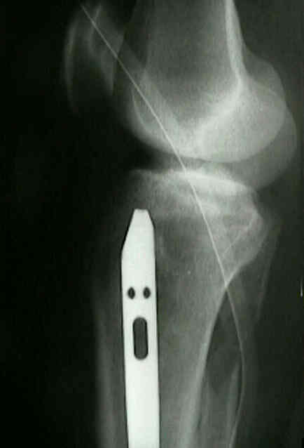
- determine nail length:
- this is best determined once the initial starting reamer is placed down the canal;
- w/ the reamer down the canal, the fracture can usually be reduced no shortening or angulation;
- use flouro to mark proximal entry position at level of tibial plateau, & distal position at physeal scar;
- radiolucent ruler:
- measures the proper nail distance;
- remember that if the ruler is placed on top of the tibia (rather beside it), then there will be a tendency to undersize length;
- pitfalls:
- do not measure nail length from the proximal tibial joint line but instead measure length from the nail entry site;
- oblique fractures tend to be shortened at the fracture site and transverse fractures or comminuted frx may be distracted;
- hence measurement of oblique fractures may cause nails to be short and transverse/comminuted fractures may cause
nails to be too long;
- with distal oblique fractures often nail ends up being too short;
- note that a nail that is too long may have the effect of distracting the fracture site, when the tip of the nail engages the
physeal scar continued impaction may distract the fracture site;
- references:
- An easy and accurate preoperative method for determining tibial nail lengths
- Tibial tubercle-medial malleolar distance in determining tibial nail length.
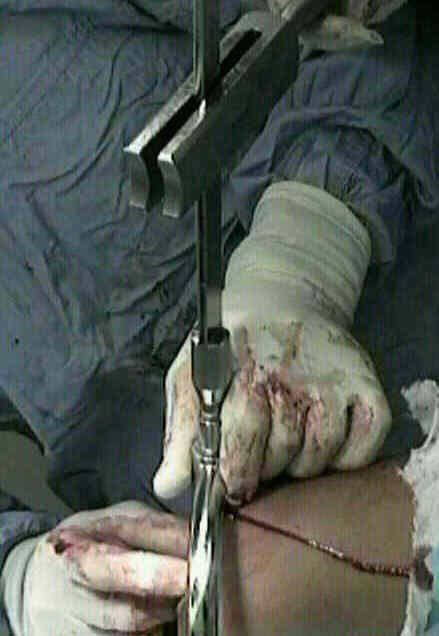
- Insertion of Nail:
- see considerations in proximal tibial fractures
- prior to nail insertion, test the proximal interlocking device to ensure that the drill will pass w/o difficulty;
- do not allow the nails to touch the skin as it is being inserted.
- check the nail progression:
- as the nail is hammered down the canal, its progression needs to be followed on the lateral view;
- if nail fails to advance with each blow of the hammer, stop, for the nail is impinging on the cortex or it too large for the canal;
- attempts to drive it further may fracture the cortex;
- although the nail may occassionally too large, the usual cause for impingement is improper alignment of the nail within canal;
- Fracture reduction: (completed as soon as the nail crosses the fracture site);
- lack of cortical continuity (fracture gap) is the one major factor that surgeon can control with regard to frx healing; (non-union)
- open fractures and transverse fractures are other risk factors for fracture non union;
- w/ proximal or distal fractures, consider need for blocking screws;
- references:
- The use of Poller screws as blocking screws in stabilising tibial fractures treated with small diameter intramedullary nails.
- Fractures of the proximal third of the tibial shaft treated with intramedullary nails and blocking screws.
- The mechanical effect of blocking screws ("Poller screws") in stabilizing tibia fractures with short proximal or distal fragments after insertion of small-diameter intramedullary nails.
- Technique for precise placement of poller screws with intramedullary nailing of metaphyseal fractures of femur and tibia.
- The logic and clinical applications of blocking screws
- [Effect of blocking screws on breakage of interlocking intramedullary nails]
- need to open the fracture site:
- Does open reduction increase the chance of infection during intramedullary nailing of closed tibial shaft fractures?
- references:
- Percutaneous cerclage wiring and interlocking nailing for treatment of torsional fractures of the tibia
- Percutaneous cerclage wiring-assisted interlocking nailing for torsional tibia fractures: a modification with improved safety and simplicity.
- Intramedullary nailing of proximal and distal one-third tibial shaft fractures with intraoperative two-pin external fixation
- Predictors of reoperation following operative management of fractures of the tibial shaft
- Percutaneous clamping of spiral and oblique fractures of the tibial shaft: a safe and effective reduction aid during intramedullary nailing..
- rotational alignment:
- once the nail crosses the fracture site, great care must be taken to restore rotational alignment;
- use flouro or the bi-malleolar axis to determine proper alignment;
- Getting the Rotation Right: Techniques for Assessing Rotation in Intramedullary Tibial and Femoral Nailing
- Findings related to rotational malalignment in tibial fractures treated with reamed intramedullary nailing
- nail centralization:
- as the nail is driven thru the proximal fragment, it is important that it centralizes prior to reaching the fracture site;
- if nail does not centralize prior to reaching fracture site, then remove nail, re-ream the canal, and consider adding a
posterior bicortical blocking screw;
- fracture compression:
- once the nail is across the fracture site, place counter pressure across the foot to provide frx compression as the nail is
driven distally;
- note that fracture compression is mainly required with mid shaft fractures, where as in contrast, distal third fractures need
to be brought out to length and held with two proximal and two distal interlocking screws;


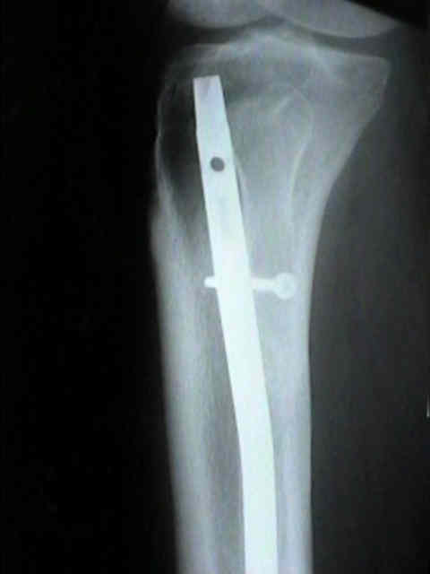
- Interlocking:
- proximal inter-locking
- distal interlocking: prior to distal interlocking, ensure that there is optimal frx site compression;
- two screws should be used if the position of the screws is within 4 cm from the fracture site;
- references:
- Single or double distal locking in intramedullary nailing of tibial shaft fractures: a prospective randomized study
- Dynamisation and early weight-bearing in tibial reamed intramedullary nailing: Its safety and effect on fracture union.
- Intramedullary nailing without interlocking screws for femoral and tibial shaft fractures
- Post Operative Care:
- if stability of the fracture is in question, then below knee cast immobilization and touch down wt bearing are used until healing;
- once partial fracture healing has taken place, consider a functional brace or consider a below knee cast with the dorsum of the
foot and ankle removed to allow ankle dorsiflexion;
- active dorsiflexion and plantarflexion stresses the tibia and produces displacements similar to wt bearing;
- references:
- Can Tibial Shaft Fractures Bear Weight Following Intramedullary Nailing? A Randomized Controlled Trial.
- static locking: most tibial fractures heal in the static locked mode;
- dynamization:
- removal of proximal or distal screws allows axial loading of tibia;
- consider at 3 months in axially stable fractures with no callus;
- axially unstable frx should remain in static mode and should receive bone grafting;
- references:
- [Intramedullary nailing of the tibia with the expert tibia nail].
- Dynamisation and early weight-bearing in tibial reamed intramedullary nailing: Its safety and effect on fracture union.
- Complications:
- ref: Prognostic Factors for Predicting Outcomes After Intramedullary Nailing of the Tibia
- non-union
- biggest risk factor for tibial non union following IM nailing is fracture mal-position or the presence of a gap at the fracture site;
- anterior knee pain:
- references:
- Anterior knee pain and thigh muscle strength after intramedullary nailing of tibial shaft fractures: a report of 40 consecutive cases.
- Incision placement for intramedullary tibial nailing: an anatomic study.
- Anterior knee pain after intramedullary nailing of fractures of the tibial shaft. A prospective, randomized study comparing two different nail-insertion techniques.
- Percutaneous intramedullary nailing of tibial shaft fractures: a new approach for prevention of anterior knee pain.
- Knee pain after intramedullary tibial nailing: its incidence, etiology, and outcome.
- Knee pain after tibial nailing.
- compartment syndrome
- reference:
- Compartment syndrome without pain!
- infection: (see infections following tibia fracture)
- if nail removal is required, then consider reaming canal after nail removal as a method of debridement;
- see addition of antibiotics to cement
- references:
- Infection after intramedullary nailing of the tibia. Incidence and protocol for management.
- Diagnosis and management of infection after tibial intramedullary nailing.
- Infection after reamed intramedullary nailing of the tibia: a case series review.
- Intramedullary infections treated with antibiotic cement rods: preliminary results in nine cases.
- The antibiotic cement nail for infection after tibial nailing.
- Antibiotic Cement-Coated Interlocking Nail for the Treatment of Infected Nonunions and Segmental Bone Defects.
- dropped hallux deformity:
- Robinson CM, et al. (1999) authors performed a prospective study of 208 patients w/ tibial frx treated by reamed IM nailing;
- 11 (5.3%) developed dysfunction of peroneal nerve, 8/11 showed a 'dropped hallux' syndrome, with weakness of EHL and
numbness in first web space, but no clinical involvement of extensor digitorum longus or tibialis anterior;
- there was good recovery of muscle function within 3-4 months in all cases, but after one year 3 patients still had some
residual tightness of EHL, and two some numbness in the first web space.
- references:
- Dropped hallux after the intramedullary nailing of tibial fractures.
- Radiographic analysis of tibial fracture malalignment following intramedullary nailing.
- Severe heterotopic bone formation in the knee after tibial intramedullary nailing.
Intramedullary nailing of femoral and tibial shaft fractures