- See:
- Pediatric Elbow Injuries
- distal humeral physeal separation
- lateral condyle frx, pediatric
- supracondylar frx of humerus
- Discussion:
- third most common pediatric elbow frx (5-10%) (behind supracondylar and lateral condylar fractures);
- most common between ages of 9 and 14 years;
- mechanism of injury is valgus strain of the joint, producing traction on the medial epicondyle thru the flexor muscles;
- frx is usually extra-articular;
- about half of cases are associated w/ dislocation of the elbow;
- the other half of cases are usually mild in nature;
- anatomy of medial epicondyle:
- medial epicondyle of humerus appears at about five yrs of age & unites w/ humeral diaphysis between 18 and 20 years of age;
- medial epicondyle is apophysis & does not contribute to longitudinal growth of the humerus;
- the apophysis is normally separated from the humeral metaphysis;
- note that the apophysis may normally appear fragmented giving the false appearance of a fracture;
- forearm flexor muscles take origin from medial humeral condyle, as does medial collateral ligament;
- ulnar nerve runs in groove in posterior aspect of this epicondyle;
- diff dx:
- osteochondrosis of the medial epicondyle:
- frx of medial condyle
- because the trochlea does not ossify until age 8 yrs, frx of the medial condyle may be mistaken for frx of medial epicondyle;
- this is especially true if there is significant pain, swelling, and instability (but no dislocation);
- consider MRI if the diagnosis is in question;
- Radiographs:
- obtain internal oblique radiographs inorder to obtain a more accurate view of displacement;
- references:
- How displaced are "nondisplaced" fractures of the medial humeral epicondyle in children? Results of a three-dimensional computed tomography analysis.
- Reliability of Internal Oblique Elbow Radiographs for Measuring Displacement of Medial Epicondyle Humerus Fractures: A Cadaveric Study
- The Distal Humerus Axial View: Assessment of Displacement in Medial Epicondyle Fractures
- Non Operative Treatment:
- minimally displaced frx can be treated easily w/ elbow flexion and neutral position (or pronation) for 3 weeks;
- even slightly displaced frxs will have good results w/ is by fibrous union;
- Operative Treatment:
- indications for surgery:
- displaced fragment trapped w/ in joint which prevents reduction;
- displacement more than 5 mm is an indication for surgery (w/ more significant displacement there will be shortening of flexor mass);
- presence of ulnar neuropathy;
- valgus instability as documented by the gravity stress test (more important in dominant arm);
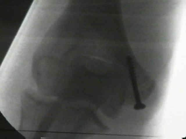
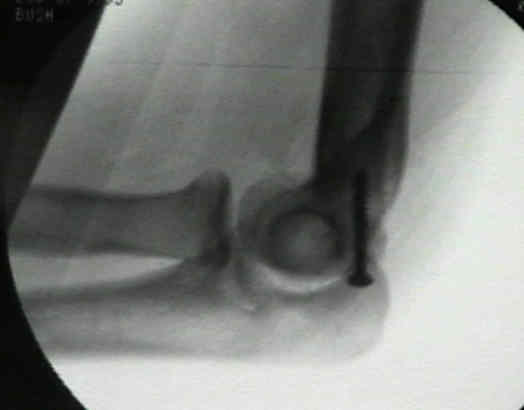
- technique considerations:
- ulnar nerve is identified and protected during exposure;
- K wires are used in young children, where as screws are used in adolescents;
- flexor pronator mass and periosteum are repaired with sutures.
- outcomes:
- Farsetti P, et al, the authors analyzed the functional and radiographic results of both nonsurgical and surgical Rx;
- 42 patients w/ displacement of >5 mm at an average age of 12 years were analyzed at avg age of 45;
- group I (19 patients):
- fracture had been treated with a long-arm plaster cast w/o reduction of the displaced medial epicondyle;
- according to a functional grading scale, there were 16 good and three fair results in Group I;
- group II (17 patients):
- ORIF with either K wires or a T-nail had been performed;
- there were fifteen good and two fair results in Group II;
- group III (6 patients):
- epicondylar fragment had been excised with suture reattachment of the tendons and the MCL;
- all but 2 patients had nonunion of fragment on follow-up radiographs, but all had a normal result on valgus stress-testing of elbow;
- ROM of the elbow was either normal or minimally decreased, and the grip strength of the ipsilateral hand was normal;
- all patients had union of medial epicondyle, w/ xray deformities of medial epicondyle, but the f(x) results were similar to those of group-I patients;
- group-III patients had four poor and two fair results;
- 4 had constant pain at the elbow and paresthesias in the distribution of the ulnar nerve;
- 1 pt had a restricted ROM of elbow, 4 patients had an unstable elbow, and three patients had decreased grip strength of ipsilateral hand;
- nonop rx of isolated frx of medial epicondyle w/ 5 - 15 mm of displacement yielded good long-term results similar to those obtained w/ ORIF;
- nonunion of epicondylar frag that was present in most patients who had been treated only w/ cast did not adversely affect functional results;
- surgical excision of the medial epicondylar fragment should be avoided because the long-term results are poor;
- ref: Long-Term Results of Treatment of Fractures of the Medial Humeral Epicondyle in Children.
- Kamath, et al.:
- operative treatment had a higher rate of osseous union than was nonoperative treatment, but it was also associated with higher rates of pain (15% vs. 8.7%) and ulnar nerve symptoms (4.5% vs. 2.5%)
- references:
- Operative versus non-operative management of pediatric medial epicondyle fractures: a systematic review
- Medial Epicondyle Fractures in Children: Clinical Decision Making in the Face of Uncertainty
Elbow dislocation with avulsion of the medial humeral epicondyle.
Treatment of fractures of the medial epicondyle of the humerus.
Management of severely displaced medial epicondyle fractures.
Operative treatment of medial epicondyle fractures in children.
Epicondylar elbow fracture in children. 35-year follow-up of 56 unreduced cases.
Deformity after internal fixation of fracture separation of the medial epicondyle of the humerus.
Operative fixation of medial humeral epicondyle fracture nonunion in children
Treatment of Medial Epicondyle Fracture without Associated Elbow Dislocation in Older Children and Adolescents
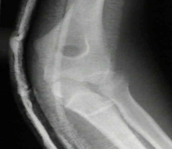
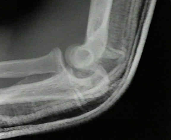
- Case Example:
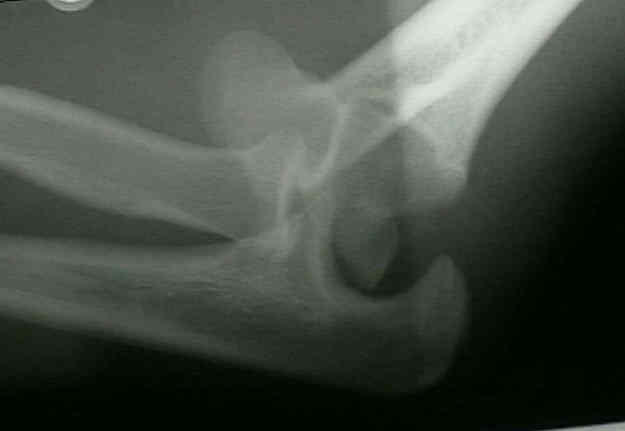
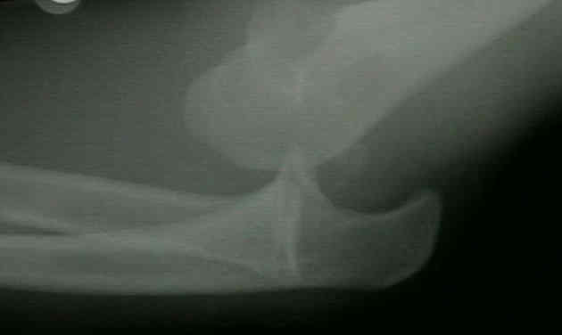

- following closed reduction, the medial epicondyle fracture is classified with regard to displacement;
- if it displaced < 5 mm and does not move w/ gentle valgus stress test, then continued closed treatment is indicated;
- ORIF of frx is indicated for displacement > 10 mm, severe valgus instability (suggested by positive gravity stress test), associated ulnar nerve sx, or incarceration of frag w/in ulnohumeral joint;