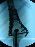
- Adult Condylar Fractures
- Pediatric Elbow Injuries
- Discussion:
- in children, supracondylar frxs typically remains extra-articular & involves thin bone between coronoid fossa & olecranon fossa of
distal humerus;
- frx line angles from anterior distal point to posterior prox site; 
- in adults, supracondylar frx of humerus may be intra-articular;
- frx occurs most often around age 6-7 years;
- classification:
- 2 types: extension type (95%) & flexion type;
- gartland classification for extension fractures:
- recognizes that anterior cortex fails first w/ resultant posterior displacement of distal fragment;
- type I: non-displaced frx;
- type II: displaced with intact posterior cortex;
- type III: displaced with no cortical contact;
- references: The prognostic value of the fracture level in the treatment of Gartland type III supracondylar humeral fracture in children.
- associated injuries:
- palpate distal radius for frx (occurs in 5-6%);
- references:
- Supracondylar elbow fractures with impaction of the medial condyle in children.
- Ipsilateral proximal metaphyseal and flexion supracondylar humerus fractures with an associated olecranon avulsion fracture.
- Ipsilateral supracondylar fracture of humerus and forearm bones in children.
- Simultaneous ipsilateral fractures of the arm and forearm in children.
- Physical Exam:
- Vascular Injuries
- Neurologic Deficits
- note that a median nerve palsy, may mask a pending compartment syndrome;
-
- Radiographs:
- posterolateral fracture displacement is correlated with median nerve and vascular compromise;
- posteromedial fracture displacement is strongly correlated with radial nerve injury
- ref: Neurovascular injuries in type III humeral supracondylar fractures in children.
- Treatment of Displaced Frx: (see type II and type III))
- it is essential to distinguish extension type from flexion type injuries;
- suspected extension-type supracondylar frx are initially splinted in 20 deg of elbow flexion pending evaluation & treatment.
- if pulse of affected arm is slightly decreased (i.e., vascular injury is a concern), then apply a continuous pulse ox so the nurses
can follow an objective measurement of perfusion;
- timing:
- if patient has recently eaten, then it may be safe to wait 8 hours to let to stomach clear its contents (this assumes that a good
pulse is present and the compartments are soft);
- Mehlman, et al. evaluated complications from early surgical treatment (< 8 hours) and delayed treatment (> 8 hours) of
displaced supracondylar frx;
- 52 patients had early treatment and 146 patients had delayed surgical treatment of a displaced supracondylar frx;
- periop complication rates of the two groups were compared with the use of bivariate and multivariate statistical methods;
- no significant difference between the 2 groups: w/ need for formal ORIF (p = 0.56), pin-track infection (p = 0.12), or
iatrogenic nerve injury (p = 0.72).
- no compartment syndromes occurred in either group;
- there were no sig difference, w/ regard to perioperative complication rates, between early and delayed treatment of
displaced supracondylar frx.
- references:
- The effect of surgical timing on perioperative complications of treatment of supracondylar humeral fractures in children.
- Effect of surgical delay on perioperative complications and need for open reduction in supracondylar humerus fractures in children.
- Early versus delayed reduction and pinning of type III displaced supracondylar fractures of the humerus in children: a comparative study.
- Delaying treatment of supracondylar fractures in children: has the pendulum swung too far?
- The effects of surgical delay on the outcome of pediatric supracondylar humeral fractures
- Outcomes of Reduction More Than 7 Days After Injury in Supracondylar Humeral Fractures in Children
- Displaced supracondylar humeral fractures: influence of delay of surgery on the incidence of open reduction, complications and outcome.
- positioning:
- references:
- Reduction and pinning of pediatric supracondylar humerus fractures in the prone position.
- reduction
- note that attempts in the ER at partial reduction and delays in reduction will only lead to increase soft tissue swelling which
will complicate the definative redection under GEA in the OR;
- percutaneous pin fixation:
- indicated for some type II frx and most type III frx;
- open reduction:
- indicated w/ vascular injuries or when closed reduction fails to achieve adequate alignment;
- postop: full return of ROM may not return for up to 1 year;
- references:
- Prospective Longitudinal Evaluation of Elbow Motion Following Pediatric Supracondylar Humeral Fractures
- Immobilization after pinning of supracondylar distal humerus fractures in children: use of the A-frame cast.
- The long-term outcome of childhood supracondylar humeral fractures: A population-based follow up study with a minimum follow up of ten years and normal matched comparisons.
- Complications:
- Cubitus Varus
- Volkmann's Contracture
- Factors affecting forearm compartment pressures in children with supracondylar fractures of the humerus.
- Vascular Injuries
- Neurologic Deficits
The Treatment of Displaced Supracondylar Humerus Fractures: Evidence-based Guideline
Supracondylar fractures of the humerus in children. Analysis at maturity of fifty-three patients treated conservatively.
Supracondylar fractures of the humerus in children: analysis of the results in 142 patients.
Tips of the trade #30. Constructing a bracket for fixation of supracondylar fractures in children.
Supracondylar fractures of the humerus: the role of dynamic factors in prevention of deformity.
Supracondylar fractures of the humerus in children.

