
 - See:
- Plating Techniques
- Monteggia Fractures in Children
- Discussion:
- Giovanni Monteggia (1814) first described frx of proximal 1/3 of ulna in association w/
anterior dislocation of radial head;
- hence dislocation of radial head w/ frx of proximal 1/3 of ulna is known as Monteggia's deformity.
- Mechanism:
- proposed mechanisms include direct blow & hyperpronation injuries as well- as the
hyperextension theory;
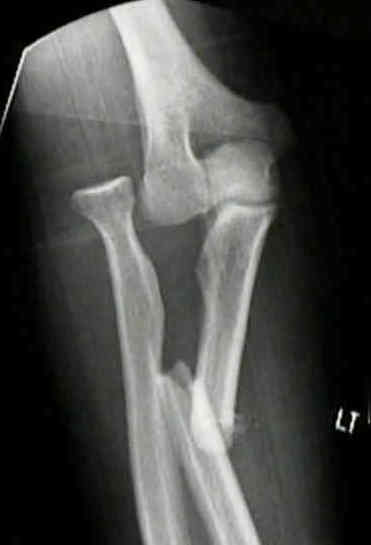
- Type I (or extension type) - 60% of cases:
- anterior dislocation of radial head (or frx) and fracture of ulnar diaphysis at any level w/
anterior angulation (usually proximal third);
- exam:
- attempt to palpate radial head (ant, post, or lateral);
- PIN palsy is most common in type I frx and may occur in a delayed fashion if the radial
head is not promptly reduced;
- reduction:
- achieved w/ forarm in full supination, & longitudinal traction;
- then elbow is gently flexed to > 90 deg to relax biceps;
- radial head is gently repositioned by direct manual pressure anteriorly on the bone;
- following reduction, radial head will be stable if left in flexion;
- angulated ulnar shaft is reduced by firm manual pressure;
- Type II (flexion type) - 15%
- posterior or posterolateral dislocation of radial head (or frx);
- frx of proximal ulnar diaphysis with posterior angulation;
- posterior Monteggia frx is reduced by applying traction to forearm w/ the forearm in full extension;
- immobilization is continued until there is union of the ulna;
- this ordinarily requires 6-10 wks depending on the age of pt;
- ref: Repair of Bado II Monteggia Fracture: Case Presentation and Surgical Technique.
- Type III - 20%
- lateral or anterolateral dislocation of the radial head;
- fracture of ulnar metaphysis;
- frx of ulna just distal to coronoid process w/ lateral dislocation of radial head;
- Type IV (5%)
- anterior dislocation of the radial head;
- frx of proximal 1/3 of radius & frx of ulna at the same level;
- Exam:
- r/o tear of the annular ligament
- associated nerve injury:
- paralysis of deep branch of radial nerve is most common;
- posterior interosseous nerve may be wrapped around neck of radius, preventing reduction;
- note: that patients whose operative treatment is delayed may be found to have a progressive PIN palsy from
constant pressure exerted by the dislocated radial head;
- spontaneous recovery is usual & exploration is not indicated;
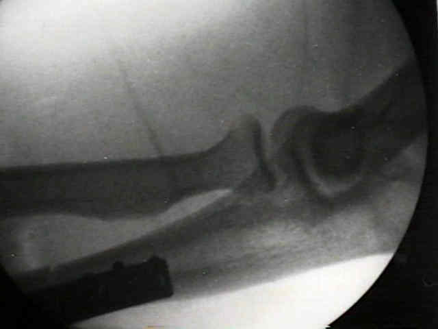
- Radiographs:
- dislocation of radial head may be missed, eventhough frx of ulna is obvious (need AP, lateral and olbique X-rays of elbow)
- line drawn thru radial shaft and radial head should align w/ capitellum in any position if the radial head is in normal position
- this is esp true on the lateral projection;
- apex of angular deformity of ulna usually indicates direction of radial head dislocation;
- Reduction:
- immobilize forearm in neutral rotation w/ slight supination, w/ cast carefully molded over lateral side of ulna at level of fracture;
- keep elbow flexed ( > 90 deg), to relax biceps, so that full supination can be avoided w/o losing reduction;
- Non Operative Treatment:
- realize that even w/ successful closed reduction of the ulna (and accompanying reduction of the radial head) that subsequently
there may be slow and progressive shortening and angulation;
- hence, these patients will require close follow up;
- Treatment:
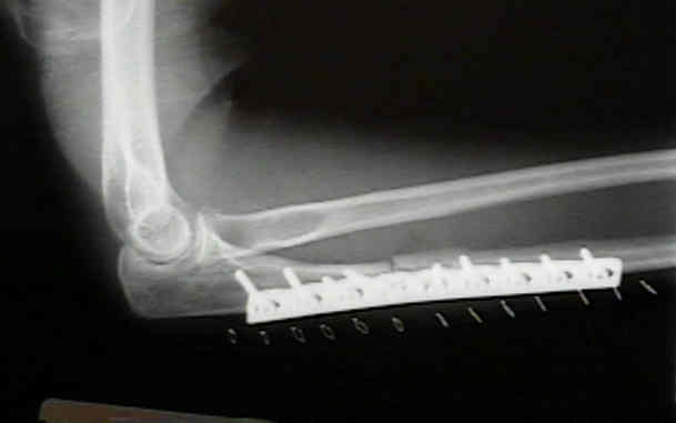
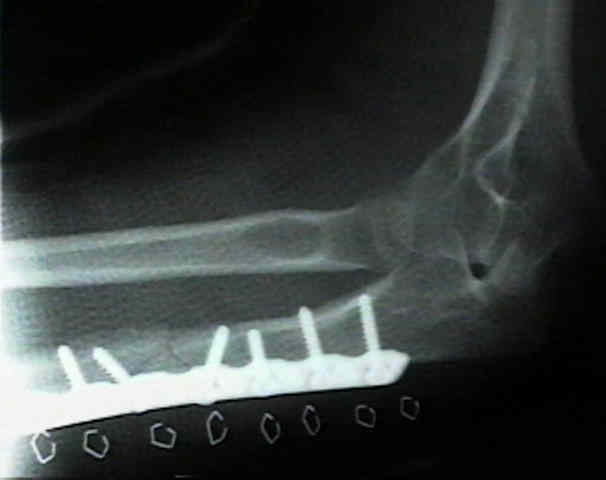
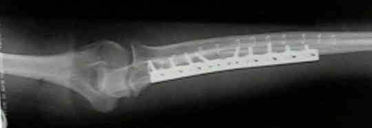
- treated by reduction and stabilization of ulna followed by reduction of radial head via supination & direct pressure;
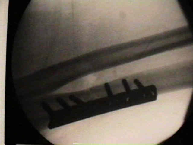
- ulnar frx is treated w/ compression plate (esp in proximal third)
- medullary nail in this location may not fill the canal and may thus provide less than rigid fixation;
- key is to obtain length and alignment, which then allows the radial head to be reduced;
- type I, III, and IV lesions are held in 110 deg. of flexion;
- type II lesions with posterior dislocations should be maintained in about 70 deg. of flexion for 6 weeks;
- Delayed Dx:
- when dx is delayed < 3 months, ORIF is indicated;
- when > 3 months has elapsed, consider non op treatment because bony ankylosis of the elbow may occur following surgery;
- bony ankylosis may be more disabling than the joint instability
- in child, a dislocated radial head should never be resected, since it will cause cubitus valgus, prominence of distal end of ulna,
and radial deviation of head;
- Complications:
- PIN or radial nerve palsy from anterior displacement of radial head;
- spontaneous recovery is usual & exploration is not indicated;
- see: nerve injuries
- non union of frx of ulnar shaft
- radiohumeral ankylosis
- radioulnar synostosis
- recurrent radial head dislocation
- myositis ossificans
- References:
The challenge of Monteggia-like lesions of the elbow mid-term results of 46 cases
Giovanni Battista Monteggia (1762-1815).
Unstable fracture-dislocations of the forearm (Monteggia and Galeazzi lesions)
Monteggia lesions in children and adults: an analysis of etiology and long-term results of treatment.
Removal of forearm plates. A review of the complications.
The posterior Monteggia lesion.
Monteggia fractures in adults: long-term results and prognostic factors
Monteggia fractures in adults.
Does a Monteggia variant lesion result in a poor functional outcome?: A retrospective study.