
- See: Soft Tissue Tumor Menu
- Discussion:
- 4th most common soft-tissue sarcoma (5 to 10 percent of all cases of soft-tissue sarcoma)
- tumor derived from the synovial tissues found along fascial planes, periarticular structures, and rarely, in joints;
- it may involve the sheaths and bursae of the tendons;
- presentation:
- most often occurs in adolescents and young adults;
- deep, painless soft-tissue mass greater than 5 cm in size is suspicious for a sarcoma;
- slowly enlarging, painless juxtaarticular mass is primary manifestation;
- usually presents as stage IIb lesion in lower limbs;
- may presents as stage I tumor in hands or feet where it may be confused with a ganglion;
- cytogenetic translocation: (X; 18) (p11; q11)
- synovial sarcomas stain positively for keratin
- location:
- typically arises in the legs and knee (knee is the most common location);
- often close to a joint;
- in the upper extremity, it is found more commonly on palmar surface;
- evidence of regional lymph node involvement strongly supports the dx;
- although synovial cell sarcomas are rare, those that do occur are frequently seen in the foot (12-18%);
- despite the name, these tumors are usually not intra-articular;
- X-rays: (see: calcification of soft tissue);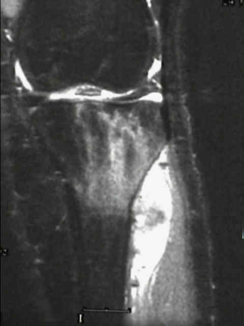
- hazy, soft tissue density w/ discrete intrinsic calcifications in 30% of cases;
- irregular contours differentiate synovial sarcoma from the phleboliths found in a benign hemangioma.
- periosteal reaction or even bone erosion or invasion;
- differential diagnosis:
- spotty calcification may indicate chondroma or hemangioma
- Bone Scans: marked radioisotope uptake;
- MRI:
- lesion is often adjacent to major neurovascular structures;
- may involve cyst formation (20%)
- Histology:
- two forms: biphasic (epitheloid) and monophasic (spindle cell type);
- biphasic form is composed of both epithelial-cell and spindle-cell components, where as monophasic form can be either epithelial-cell or spindle-cell type;
- spindle-cell form:
- predominate spindle cell component (monophasic synovioma) contains cords of spindle cells which may resemble fibrosarcoma;
- diff dx:
- malignant hemangiopericytoma
- fibrosarcoma
- spindle-cell squamous-cell carcinoma;
- epithelioid (w/ glandular component):
- might be confused with adenocarcinoma;
- reveals a biphasic pattern: intermixed areas of "glandular" synovial like cells & spindle shaped fibrous cells;
- this glandular area will stain PAS positive;
- synovial cells:
- have an acinar, ductal, or longitudinally arranged tall columnar cells around acellular "slits" containing mucin;
- fibrous component:
- arranged in the herringbone pattern of fibrosarcoma;
- Prognosis:
- recurrence rate is high
- lesion characteristically metastasizes to lymph nodes, bones, and lungs;
- other sarcomas which spread via lymph nodes include: clear cell sarcoma, epithelioid sarcoma, rhabdomyosarcoma
- 5-year survival rate ranges from 25 to 55 %;
- Treatment
- low grade lesions: treated w/ wide excision;
- high grade tumors:
- requires either radical resection or wide surgical excision plus XRT;
- if tumors are greater than 8 cm in diameter then consider administering chemotherapy and radiation therapy;
- radiation therapy may provide local control and can create a pseudocapsule around the tumor;
- use chemotherapy if there is metastatic disease
Synovial sarcoma.
Tendosynovial sarcoma: a clinicopathological study of 136 cases.
Soft tissue tumors of the foot and ankle.
SYT-SSX gene fusion as a determinant of morphology and prognosis in synovial sarcoma.