- Anesthesia:
- GEA if possible (a neurological exam is often required at the end of the case);
- Equipement:
- bipolar cautery
- hand tray
- bone reduction clamps
- small Homans
- small fragment set (Synthes - Allen Burrus - # 919 981 1204)
- 0.45 K wires
- flouro
- roungeurs
- 5-0 chromic sutures
- 0-Vicryl sutures
- thyroid or similar retractor
- Prep:
- need to prep out for bone graft (contra-lateral iliac crest)
- Surgical Approach: 
- incision starts at thenar crease of palm, curves toward middle of forearm, w/ transverse segment as it crosses volar flexion crease of wrist;
- FCR tendon is then retracted ulnarly and the incision is continued thru dorsal sheath, down to pronator quadratus muscle;
- further exposure can be achieved by extending the incision along the border of the thenar crease, and by releasing the FCR from its attachement to the trapezium;
- alternatively, the incision is carried down between the FCR and radial artery;
- radial artery need not be exposed;
- it is simply retracted radially and protected by the surrounding soft tissues;
- w/ CTS symptoms use standard open approach, but do not extend the incision to the more proximal wound (inorder that injury to the palmar cutaneous branch is avoided);
- pronator quadratus muscle is taken down from its radial origin to expose the underlying fracture;
- this will need to be repaired at the end of the case;
- since repair of the pronator can be difficult, it can be tied down to the radial edge of the butress plate;
- avoid excessive retraction of the median nerve;
- reduction:
- reduction is achieved with supination and dorsiflexion over rolled towel, and is confirmed w/ flouroscopy;
- reduction is held w/ one or two K wires inserted from volar to dorsal, which are oriented to allow the holes of the T plate to slide down the wire;
- volar capsule should not be opened for inspection for this would violate important radioscaphocapitate and radioscapholunate ligaments;
- when arthrotomy is needed, it is best performed dorsally between third (EPL) and fourth (EDC) dorsal compartments, which provide superior view;
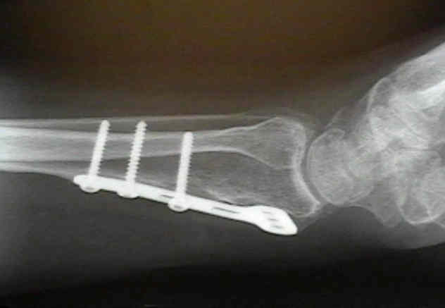
- articular reconstruction is supported by autogenous cancellous-bone graft, as well as a small buttress plate;
- Implants:
- volar plates are well tolerated, and seldom need to be removed;
- small T plate on the volar aspect;
- plate is bent in mid portion to effect prebending effect;
- ensure that the distal margin of the plate does not encroach on the articular surface (using flouro);
- slight dorsal articular penetration may be allowable;
- begin w/ a proximal screw (3.5 cortical) placed into the distal side of the oval hole, which will have the effect of moving the plate distally (which then applies compression to the periarticular fragments);
- subsequently, insert the distal screw (4.0 cancellous) only if it is needed