- See:
- Shoulder Arthrography
- MRI
- Impingement Radiographic Series:
- axillary view: may reveal an Os Acromiale, which is associated w/ impingment;
- scapular outlet view
- allows assessment of acromial morphology;
- examination of cadavera reveal:
- type 1, a flat acromion (17% of shoulders): 3% of all cuff tears have this type of acromion;
- type 2, a curved acromion (43%): 27% of all cuff tears have this type of acromion;
- type 3, a hooked acromion (40%): majority (70 - 90%) of rotator cuff tears may be seen in pts w/ type-2 or a type-3 acromion
- type A: less than 8 mm in thickness;
- type B: 8-12 mm thick;
- type C: greater than 12 mm in thickness;
- references:
- The morphology of the acromion and its relationship to rotator cuff disease. Bigliani LU, et al. Orthop Trans. 1986;10:228.
- The clinical significance of variations in acromial morphology. Morrison DS, Bigliani LU. Orthop Trans. 1987;11:234
- A modified classification of supraspinatus outlet view based on configuration and anatomic thickness of acromion. Orthop. Trans. 767. 1992-1993.
- 30 deg Caudal Tilt AP View: is taken tangential to dome of acromion to assess size of anterior inferior acromial osteophyte;
- AP of the Shoulder
- note that normal acromiohumeral interval is 1 to 1.5 cm;
- other varients of the AP view is:
- internal rotation view;
- 35 deg external rotation;
- 90 deg abduction view;
- Grashey view:
- obtained w/ 30 deg lateral oblique projection, tangential to glenohumeral joint, in order to obtain view directly down joint to reveal any degenerative changes;
- Active Abduction View
- West Point View: may be indicated in younger patients w/ suspected anterior instability;
- Specific Pathologic Findings:
- AC joint arthrosis
- superior subluxation:
- active abduction view
- decreased space between humerus & inferior portion of acromion;
- usually indicates poor rotator cuff function, highly attenuated rotator cuff, or massive tear of rotator cuff;
- pts w/o rotator cuff tears or with small to medium size tears may have normal appearing plain radiographs with an acromiohumeral interval of 1 to 1.5 cm;
- when acromiohumeral interval measures < 5 mm or less, significant tear of the rotator cuff should be suspected;
- disadvantages: this view can be unreliable due to position of the arm and x-ray tube;
- osteoarthritic changes:
- prominent osteophyte at the inferior margin of the humeral head or glenoid is characteristic;
- mild arthrosis: inferior osteophyte less than 3 mm in length;
- moderate arthrosis: inferior osteophyte between 3-5 mm in length, irregularity of the joint line and subchondral sclerosis;
- severe arthrosis: inferior osteophyte measuring more than 5 mm or if there is joint incongruity;
- greater tuberosity:
- atrophy of greater tuberosity, cystic changes in anatomic neck of humerus, notching of articular surface of head & greater tuberosity, & irregular new bone formation at lateral acromion;
- in chronic rotator cuff disease, with or without tearing, there may be evidence of greater tuberosity sclerosis and spurring w/ cyst formation and subacromial spurring;
- Case Example:
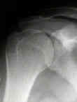
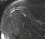
- a large 40 year old male who was felt to have isolated impingment by both clinical exam and by MRI;
- at arthroscopy, it was found that the patient had a 2 cm supraspinatus tear which was covered by a thick layer of bursa;
- the radiographs reveal sclerosis of the greater tuberosity and the MRI shows fluid in the subacromial bursa
Optimal plain film imaging of the shoulder impingement syndrome.


