
- See:
- Total Hip Replacement Menu:
- total hip in DDH
- acetabular component menu:
- crowe classification
- Anatomic Position - Discussion:
- w/ low dislocation, attempts should be made for antatomic placement, but should also be considered w/ high dysplastic dislocation;
- long term success rate should be 90% at 10 years;
- this takes advantage of better bone stock at the site of the true acetabulum;
- this may restore Shenton's line but osseous coverage may be limited (especially anteriorly);
- anatomic positioning may not be possible if the hip is stiff from scarring (from previous surgeries);
- femoral component:
- shortening procedure:
- femoral neck shortening;
- indicated when there is a need for only a few cm of shortening;
- femoral neck shortening is performed along with release of the psoas and external rotators;
- femoral neck is sequentially shortened until trial reduction is possible, and may be shortened down to lesser trochanter;
- subtrochanteric shortening:
- indicated when several cm of shortening is required or if derotational osteotomy is required;
- generally this will be necessary if there is more than 5 cm distance between the lesser trochanter and the transischial line;
- affords excellent exposure of acetabulum through retraction of the proximal fragment superiorly or anteriorly;
- performed as an oblique or step cut osteotomy;
- once the osteotomy and initial shortening are completed a trial femoral component is inserted and the hip is reduced;
- at this point change in leg length is determined and tension on the sciatic nerve is checked (further femoral shortening may be required);
- proximal fragment is then retracted superiorly or anteriorly to give exposure of the acetabular exposure and reconstruction;
- soft tissue releases are performed as needed in order to mobilize the proximal femoral fragment;
- subtrochanteric osteotomy avoids the fixation problems inherent with trochanteric osteotomy;
- acetabular component:
- either bone graft or cement is needed to fill superior defect; (see bone grafting of acetabular defects)
- as pointed out by Mackenzie, et al (1996), use of cement to fill the superior defect was 85% successful at 15 years (using revision as a criteria for failure);
- Crow III (no superior roof) required the greatest amount of bulk cement (w/ the cement comprising 100% of the component's roof);
- when reaming, direct most of the reaming in a superior and posterior direction, and take care not to fracture the anterior wall;
- sciatic nerve palsy;
- in DDH, the sciatic nerve never reached its normal adult length, and therefore the nerve is intolerant of being stretched to a "normal length;"
- this is in contrast to an adult w/ a shortened extremity due to trauma in which case the limb can often be lengthened to its original position;
- at the end of the case, it is important to check the nerve tension with the hip extended and knee flexed (relaxing the nerve) and to check the nerve
with the hip flexed and knee extended (which places the nerve under maximal tension);
- trochanteric non union:
- if a trochanteric ostoetomy has been performed, and the acetabular component has been placed in an anatomic position then the trochanter may not
reach the trochanteric bed at wound closure;
- this may require lengthening of the medius tendon or elevation from the innominate bone;
- references:
- Treatment of high dislocation of the hip in adults with total hip arthroplasty. Operative technique and long-term clinical results.
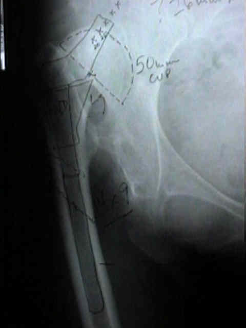
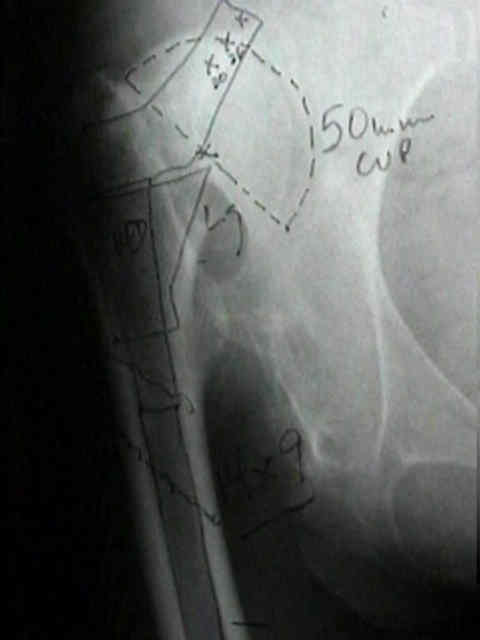
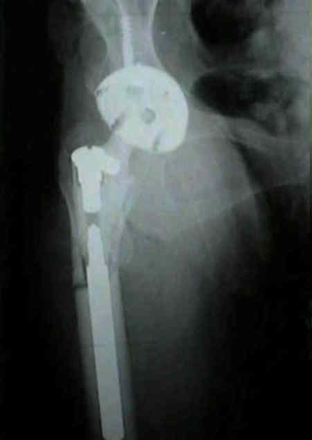
- case example:
Coxarthrosis after congenital dysplasia. Treatment by total hip arthroplasty without acetabular bone-grafting.
Arthroplasty in high congenital dislocation. 21 hips with minimum five-year follow-up.
Total hip replacement for coxarthrosis secondary to congenital dysplasia and dislocation of the hip. Long Term Results.
Total Hip Arthroplasty with Bulk Femoral Head Autograft for Acetabular Reconstruction in Developmental Dysplasia of the Hip