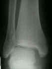
- Discussion:
- w/ an "isolated fibular frx," the paramount indication of an unstable frx is lateral talar shift (which indicates deltoid ligament injury);
- this is manifested radiographically as an increase in the medial clear space;
- normally the medial clear space (between talus & medial malleolus) should be less than 4 mm, and when the medial space is > than 4 mm, then lateral shift of talus is present;
- AP view:
- often, it is easier to measure medial clear space on AP view rather than mortise view due to obliquity of the medial mortise;
- width of medial clear space should equal the superio-medial clear space;
- disruption of deltoid ligament is present when medial clear space is increased > 3-5 mm;
- mortise view:
- due to obliquity of medial malleolus, measurement of medial clear space can be difficult on mortise view (in this case use AP view);
- attempt to use similar AP borders for measurements;
- anterior edge of medial malleolus to anterior talus
- posterior edge of medial malleolus to posterior talus;
- superior joint space should be within 2 mm medially of its width laterally;
- because xray measurements can vary (due to technique and magnification), it is also useful to look at general symmetry of the mortise;
- normally there is symmetric radiolucent line around the talus of equal width;
- when it is difficult to judge whether medial clear space widening is present, draw a line down the center of the tibial shaft, and a line down the center of the talus;
- normally the two lines should be colinear, and any overlap indicates lateral talar shift;
- Cautions:
- w/ isolated lateral malleolar fractures, an impression of lateral shift may result, in part, from external rotation of talus and ankle plantar flexion (which together, profiles the more narrow posterior part of the talus);
- the result give a false impression of a widened medial clear space
Radiographic Measurement of the Distal Tibiofibular Syndesmosis Has Limited Use.
Radiographic Measurements Do Not Predict Syndesmotic Injury in Ankle Fractures: An MRI Study.
Understanding the superior clear space in the adult ankle.
Does a Positive Ankle Stress Test Indicate the Need for Operative Treatment After Lateral Malleolus Fracture? A Preliminary Report.
Comparison of manual and gravity stress radiographs for the evaluation of supination-external rotation fibular fractures.
Stress radiographs after ankle fracture: the effect of ankle position and deltoid ligament status on medial clear space measurements.
Ankle stress test for predicting the need for surgical fixation of isolated fibular fractures.
Stress examination of supination external rotation-type fibular fractures.
Plantar Flexion Influences Radiographic Measurements of the Ankle Mortise

