
- See:
- Q angle
- Malalignment of Patella
- Discussion:
- distal realignment procedures are chiefly indicated in pts w/ patellar subluxation, absence of hyper-laxity, and increased Q angle;
- as pointed out by Fulkerson, et al (1990), less than 2% of patients w/ patellofemoral pain will require tibial tubercle transfer;
- as noted by Morshuis, et al, distal realignment procedures result in satisfactory results in about 2/3 patients who have patellofemoral pain and x-ray evidence of arthrosis but nearly all patients w/ patellofemoral pain w/o x-ray evidence of arthrosis had good or excellent results;
- note that distal realignment procedures are generally only indicated in skeletally mature patients;
- Clinical Findings:
- subluxation of the patella
- malalignment of patella
- Q angle
- movie sign: retro-patellar pain due to prolonged passive knee flexion;
- Disorders of patellofemoral alignment.
- Anteromedialization of the tibial tuberosity in the treatment of patellofemoral pain and malalignment.
- Radiographic Findings
- Fulkerson Osteotomy:
- modification of the Elmslie-Trillat Procedure, but involves anterior displacement as well;
- main indications are persistent pain and moderate articular degeneration;
- allows anteriorization of upto 15 mm, which should decrease lateral facet contact pressure;
- arthroscopy should procede the osteotomy to document patellar arthrosis;
- incision and exposure:
- lateral retinacular release: linear incision allows for concomitant lateral retinacular release, if lateral tilt is also present;
- references:
- Improvement of proximal tibial osteotomy results by lateral retinacular release.
- Lateral patellar retinaculum tension in patellar instability.
- incision is made just lateral to the patella, across the tibial tuberosity, and further distally along the anterior ridge of the tibia;
- carefully elevate the anterior compartment musculature from the lateral surface of the tibia;
- note that the deep peroneal nerve and the anterior tibial artery lie on the interosseous membrane, near the posterior cortical surface;
- carefully delineate the medial and lateral borders of the patellar tendon and tuberosity;
- plane of the osteotomy:
- incise through the periosteum along the medial side of the tuberosity a distance 5-8 cm distal to the tuberosity;
- if anteriorization is required (requiring a steeper osteotomy), then make periosteal incision closer to the tibial crest;
- distally the osteotomy should narrow and taper which allows it to act as a pivot for the more proximal portion of the tuberosity;
- insert K wires from the anteromedial tibial surface to the posterolateral surface;
- each drill should be seen to exit the posterolateral tibial surface;
- if 1.25 mm of anteriorization are needed, then the osteotomy plane is made steep, however, the osteotomy should
not encroach on the posterior tibial cortex, since this risks injury to the posteriorl neurovascular structures;
- as noted by Ferguson, et al, elevation of 1.25 cm decreases patellofemoral contact forces by 60-80%, but further elevation provided less benefit;
- usually no more than 1 cm of medialization is needed;
- osteotomy cut:
- typically a 5-8 cm bone pedicle will be require for adequate bone healing;
- medial osteotomy cut should follow the plane of the K wires (from antero-medial to postero-lateral);
- lateral osteotomy cut is required at the most proximal part of the osteotomy and must be directed anteriorly to prevent unexpected propagation of the osteotomy;
- care is taken to leave an intact distal taper of bone, which serves as a hinge;
- note that the deep peroneal nerve and the anterior tibial artery lie on the interosseous membrane, near the posterior cortical surface;
- references:
- Elevation of the insertion of the patellar ligament for patellofemoral pain
- Biomechanical analysis of flat and oblique tibial tubercle osteotomy for recurrent patellar instability.
- position the tuberosity:
- determine the position of the tuberosity which allows the best excursion of patella in the intercondylar groove;
- usually no more than 1 cm of medialization is needed;
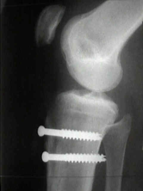
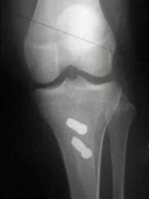
- fixation:
- fixation is carried out w/ 4.5 cortical screw
- the bone tap should be carefully applied to avoid neurovascular injury;
- often two bone screws are needed;
- as noted by Morshuis, et al, 24 out of 25 patients had pain over the screw site, which required subsequent screw removal;
- Anteromedialization of the tibial tuberosity in the treatment of patellofemoral pain and malalignment.
- complications:
- pain over the screw site in most patients;
- loss of ability to kneel down;
- proximal tibial fracture
- injury to popliteal artery and its trifurcation;
- references:
- Fracture of the proximal tibia six months after Fulkerson osteotomy. A report of two cases.
- Fracture of the proximal tibia after Fulkerson anteromedial tibial tubercle transfer. A report of four cases.
- Fracture of the proximal tibia with immediate weightbearing after a Fulkerson osteotomy.
- Vascular risk associated with bicortical tibial drilling during anteromedial tibial tubercle transfer.
- references:
Anteromedial tibial tubercle transfer without bone graft.
Anteromedialization of the tibial tuberosity for patellofemoral malalignment.
- Historical Operations:
- Hauser Procedure:
- discussed for historical purposes only;
- involves medialization of the tibial tubercle inorder to decrease Q angle;
- due to the anatomy of the proximal tibia, translating the tibial tubercle medially, will also translate the tubercle posteriorly;
- posterior translation of the tibial tubercle will have the effect of increasing patellofemoral contact pressures which leads to DJD;
- reference:
- Total tendon transplant for slipping patella: a new operation for recurrent dislocation of the patella. 1938.
- Maquet Procedure:
- discussed for historical purposes only;
- involves anterior translation of the tibial tubercle which has the effect of decreasing patellofemoral contact forces;
- patients w/ pain due to early patellofemoral arthrosis may expect pain relief following the Maquet Procedure;
- disadvantages w/ this procedure include high incidence of skin necrosis, and no effect on the Q angle;
- references:
- Clinical assessment of Maquet tibial tuberosity advancement.
- The Maquet procedure. A retrospective review.
- The Maquet procedure in the treatment of patellofemoral osteoarthrosis. Long-term results.
- Long-term follow-up study on the Maquet procedure with special reference to the causes of failure.
- Elmslie-Trillat Procedure:
- medial tibial tubercle transfer which has no posterior displacement;
- does not involve anterior displacement of the tuberosity;
- references:
- Use of a modified Elmslie-Trillat procedure to improve abnormal patellar congruence angle.
- An evaluation of the Elmslie-Trillat procedure for management of patellar dislocations and subluxations. A preliminary report.
Biomechanical effects of different surgical procedures on the extensor mechanism of the patellofemoral joint.
Elevation of the insertion of the patellar ligament for patellofemoral pain.
The treatment of patellofemoral pain by combined rotation and elevation of the tibial tubercle.
Anteromedialization of the tibial tuberosity for patellofemoral malalignment.
A modified tibial tubercle osteotomy for patellar maltracking: results at two years..