
- See:
- Diskitis
- Gun Shot Wounds to the Spine
- Tuberculous Spondylitis
- Discussion:
- in adults, lumbar infections may often originate from urinary tract infections which drain thru Batson's venous plexus;
- respiratory system is also a source of blood borne infection;
- patients may present 1-8 weeks from the original inciting event;
- purulent material may break out of cortex anteriorly to form paravertebral abscess or posteriorly to form an epidural abscess.
- weakening of the bone may cause vertebral collapse.
- hematogenous osteomyelitis is seen more often in the child than the adult;
- since the adult does not have an epiphyseal plate, site of origin is different from that of childhood;
- risk factors:
- diabetes (upto 25%)
- older debilitated patients;
- IV drug addicts (at risk for pseudomonas infection);
- h/o of pneumonia, UTI, skin infection;
- immuno-comprimised patient;
- site of infection:
- lumbar spine is the area most often affected by spinal osteomyelitis;
- infections in this area uncommonly cause paralysis;
- thoracic & cervical regions are affected less often but have a higher incidence of paralysis in that order;
- bacterial diff dx:
- staph aureus is most common but MRSA is also common;
- also consider strep viridans;
- in drug addicts & immunocomprimised pts, diff dx includes brucellae, candidae, or coccidiomycosis;
- propionibacterium acnes:
- ref: Propionibacterium acnes Vertebral Osteomyelitis: Seek and Ye Shall Find?
- tuberculosis:
- ref: Clinical problem-solving. Beware of first impressions.
- diff dx:
- infectious discitis:
- often is caused by hematogenous spread of bacteria from an infectious focus (infected tooth or urinary tract), but direct infection may occur from an infected aortic prosthesis or rarely from a fish bone lodged in the throat;
- ref: Cervical spondylodiscitis after removal of a fishbone. A case report.
- post operative disc space infection
- neoplasm
- renal spondyloarthropathy
- tuberculous spondylitis
- fungal osteomyelitis
- Clinical Findings:
- associated with a significant delay in diagnosis (6-12 weeks)
- may have insidious course, w/ back pain developing over 1-3 mo, often w/o fever, wt loss, or any other systemic signs.
- h/o of unremmiting spine pain at any level is characteristic, & tenderness, spasm, and loss of motion are seen;
- initially onset of pain may be insidious;
- w/ time, pain may be so severe that jarring the bed may be agonizing;
- pain restricts ROM of spine, & percussion over spinous process is tender;
- children with vertebral osteomyelitis are systemically ill (unlike diskitis);
- symptoms include fever and an increased leukocyte count;
- hip joint: w/ acute hip pain, flexion contracture, & limited motion;
- abdominal syndrome: w/ sx & signs that may suggest acute appendicitis;
- meningeal syndrome;
- paralysis:
- incidence of neurological deficit may be as high as 40%;
- onset of paralysis may suggest epidural extension from abscess;
- abscess is usually located anterior to neural elements;
- associatted conditions:
- older age
- higher vertebral level of infection (cervical)
- presence of debilitating dz such as diabetes, R.A., steroid use;
- staph aureus infections;
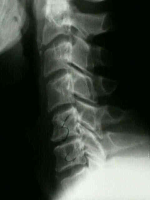
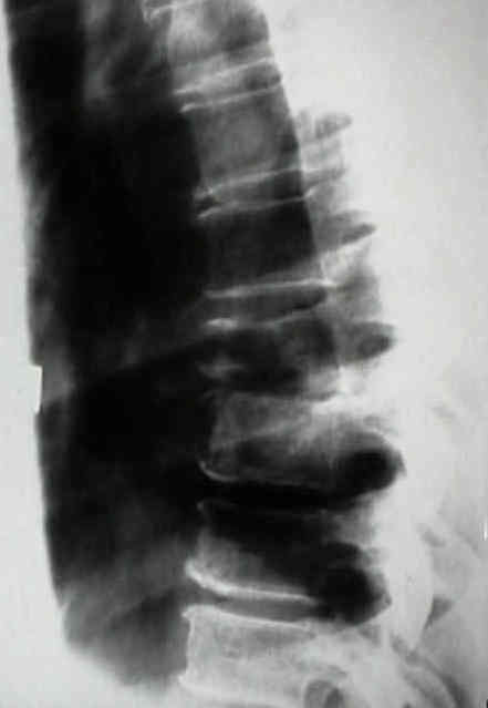
- Radiographic Features:
- erodes vertebral body endplate;
- osteolysis occurs most often but marginal scleroses of varying depth may occur due to trabecular collapse
and new bone formation;
- rarefaction of adjacent vertebral bodies is noted at 6 weeks;
- reactive screrosis at 8 weeks, & new bone formation is noted at 12 wks;;
- intervertebral fusion is usally signifies a resolution of process usually occurring after 6 mo and 2 years;
- at 2 weeks, disc space narrowing occurs;
- usually neoplasms will not narrowing of disc space;
- infections involving vertebral bodies frequently extend into & destroy adjacent intervertebral discs.
- MRI dx of Spinal Osteomylitis:
- on coronal views look for incr uptake (on T2) to evaluate for psoas abscess;
- reference:
- Neuroradiology: Review Article: Role of MR Imaging in the Management of Spinal Infections.
- Work up:
- sed rate: osteomyelitis of the spine
- determine site of infection:
- vertebral bodies, posterior elements of spine, epidural space, intervertebral disc, and the perispinal soft tissues;
- must also distinguish vertebral osteomyelitis from diskitis which occurs after surgical resection of a vertebral disc;
- usually two vertebral bodies and a disc space affected;
- consider bacterial endocarditis;
- gun shot wounds to the spine
- if missile first enters the gut prior to passing thru the spine, gut flora may be passed directly into the spinal canal;
- references:
- Infection about the spine associated with low-velocity-missile injury to the abdomen.
- differential dx:
- determined from blood cultures & percutaneous needle biopsy;
- needle bx should be performed through posterolateral approach in thoracic and lumbar spines;
- Craig needle is helpful in obtaining a subcortical biopsy;
- smaller needles are necessary for bx of C spine & L5 vertebra;
- infections of upper cervical spine & sacrum are not readily or safely accessible to needle aspiration;
- open bx is indicated when tissue dx has not been made, & anterior approaches or costotransversectomy are utilized;
- gram stain:
- gram negative bacilli
- gram negative cocci
- gram positive bacilli
- gram positive bacilli
- Non Operative Rx:
- IV antibiotics are started along w/ spinal immobilization, including bed rest, bracing, or combination of both;
- see bacterial menu:
- gallium scan can be used to follow the resolution of the disease;
- sed rate can be used to follow the resolution of the dz;
- 50% of patients will fuse spontaneously;
- prognostic signs:
- good prognostic signs for successful non operative treatment included:
- age less than 60 years;
- normal immune status;
- decreasing sed rate;
- infection w/ staph aureus
- Indications for Surgical Debridement:
- systemic signs which continue despite antibiotic treatment;
- progressive vertebral body collapse or kyphosis;
- evolving neurologic deficits:
- progressive neurologic deficit may be the result of progressive abscess formation or vertebral collapse;
- in either case, surgical debridement and stabilization will be required;
- any degree of myelopathy;
- Surgical Options:
- open biopsy, I & D, and bone grafting;
- laminectomy:
- indicated only for drainage and debridment of posterior element infections and localized epidural abscess;
- bone grafting:
- autogenous bone grafting is indicated following debridment as long as intravenous antibiotics are administered;
- anterior debridment & strut grafting:
- for involving neurolgic deterioration, bony destruction, or kyphosis:
- for drainage of anterior abscesses;
- role of spinal instrumentation in such cases is controversial;
- w/ large deformity, posterior instrumentation may be indicated;
- treatment of paralysis:
- treatment consists of IV antibiotics & early decompression, autogenous bone grafting, and appropriate spinal stabilization and/or bracing;
- nonoperative rx offers little hope for neurologic recovery;
- anterior approach provides better exposure for draining associatted psoas and paraspinal abscesses
Postoperative posterior spinal wound infections.
Pyogenic infection of the spine.
Echinococcal infestation of the spine in North America.
Pyogenic vertebral osteomyelitis with paralysis. Prognosis and treatment.
Analysis of 61 cases of vertebral osteomyelitis.
The spectrum of intervertebral disc-space infection in children.
Vertebral osteomyelitis in infants.
Images in clinical medicine. Vertebral osteomyelitis.
Pyogenic and fungal vertebral osteomyelitis with paralysis.
Pyogenic Vertebral Osteomyelitis.
Pyogenic non tuberculous spinal infection: an analysis of 30 cases.
Anterior cervical debridement and strut grafting for osteomyelitis of the cervical spine.