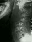
- See:
- Myobacterium tuberculosis
- Vertebral Osteomyelitis

- Discussion:
- 4000 cases of extrapulmonary involvment each year in the US;
- most common extra pulmonary location of TB is the spine;
- spine is the extrapulmonary site in over 50% of the cases of bone and joint involvement;
- about 2/3 of patients will have abnormal chest x-rays;
- severe kyphosis, sinus formation, & (Pott's) paraplegia are all late sequelae;
- sites of involvement:
- thoracolumbar spine is most common site of involvement (50% of patients);
- cervical spine and lumber spine are each involved in 25% of patients;
- pelvis, femur, and tibia may each be involved in 10% of patients;
- clinical features:
- neurologic deficit is uncommon in children;
- spinal cord injury may occur secondary to direct pressure from abscess, bony sequestra (good prognosis);
- most common presenting symptom is back pain, but systemic signs, such as fever, night sweats, anorexia, and weight loss, are common;
- associated injuries:
- probably more significant is the fact that any patient with spinal TB has 50% chance of having associated pulmonary, renal, or other viceral TB in their past medical history;
- Anatomy of Spinal Involvment
- infection may originate anteriorly in the vertebral body near anterior cortex, in the center of the body, or adjacent to the end plate;
- unlike disc space infection in children, TB affects vertebral bodies and does not destroy the disk until very late in the disease;
- originating from metaphysis of vertebral body and spreading under anterior longitudinal ligament, spinal TB can cause destruction of several continguous levels or can result in skip lesions (15%) or abscess formation (50%);
- Peridiscal - 33%
- disease begins within metaphyseal bone
- spread occurs beneath the anterior longitudinal ligament to involve adjacent areas;
- disc is spared;
- Anterior 2.1%
- disease begins and spreads beneath the anterior longitudinal ligament involving several levels;
- x-rays show anterior vertebral body scalloping;
- Central 11.6%
- disease limited to the middle of a single vertebral body;
- frequently leads to vetebral collapse w/ result kyphotic deformity
- Diagnostic Studies:
- sed rate:
- sed rate of < 50 mm / hour may indicate uncomplicated TB osteomyelitis rather than pyogenic form;
- skin testing:
- in the U.S. about 10-15% of the population will have positive test;
- w/ infection, skin tests are usually, but not always, positive;
- false negative tests will occur in malnourished patients and AIDS patients;
- skin testing in a patient w/ an active infection may result in skin slough;
- histology:
- from CT guided needle biopsy, look for granulomatous pattern w/ caseating necrosis & giant cell formation;
- acid fast bacilli on staining may or may not be seen, and cultures are frequently negative;
- note, due to the high occurance of false negative results w/ aspiration, attempt to obtain a tissue sample to assist w/ the dx (may not always be possible);
- radiographs:
- contiguous vertebral involvment w/ diffuse osteopenia, erorsions, kyphosis, and ultimately bony or fibrous briding;
- it usually involves body of vertebra, sparing the posterior elements;
- typically appearance involves anterior destruction of two adjacent vertebrae and destruction of the intervening intervertebral disc (in some cases eroded vertebrae will be at different levels);
- disc involvement will help shift the diagnosis away from malignancy and toward infection;
- note that myeloma and lymphoma may cause disc erosion;
- bone scan:
- unreliable for diagnosis of acitve TB (cold scans in upto 35-40%);
- CT scan:
- helps define the extent of soft tissue involvement inaddition to osseous destruction;
- soft tissue calcification will help distinguish spinal TB from other conditions;
- Non Operative Treatment:
- if detected early (before collapse of more than 1-2 vertebral body) treatment consists of antibiotics and immobilization;
- even w/ mild kyphosis (but no neurologic deficit), antibiotics & bracing are used;
- w/ adequate medical treatment, there may be significant resolution of neurologic symptoms, and there will be a halt in the progression of kyphosis;
- in young children, there will often be some resolution in the kyphosis, especially if only one or two vertebrae are involved;
- antibiotics for all patients at the outset, reserving surgery for cold abscesses that are palpable posteriorly, as well as for those cases w/ neurological environment that have failed to improve in response to 2-3 months of antiTB therapy and immobilization;
- 2 months of pyrazinamide, isoniazid, and rifampin given qd, followed by 4 months of INH and rifampin;
- outcomes:
- assessment of outcome should include prevalence of symptoms, amount of physical activity, amount of CNS involvement, presence/absence of sinus and/or abscess, and radiographic status of the lesion;
- in the report by the Working Party on Tuberculosis of the Spine (1998), excellent results were achieved with outpatient chemotherapy, with no late relapse or late onset paraplegia;
- with non operative therapy, there is no advantage w/ wearing a plaster jacket nor is there any advantage to bed rest;
- the only advantage of radical debridement was less late deformity;
- a short course of INH and rifampin were as effective as the standard 18 month regime (which consisted of para-aminosalicyclic acid and INH);
- in the report by Parthasarathy R, et al (1999), ambulant chemotherapy for 6 months with daily INH plus rifampicin was as effective as radical anterior resection plus antibiotics (randomized control trial with 10 year followup);
- the authors recommended that INH, rifampicin, pyrazinamide, and ethambutol, given 3 times per week for 2 months followed by INH and rifampicin given three times per week for 4 additional months;
- the main subgroup that failed medical treatment was in patients younger than 15 yrs with more than 30 deg kyphosis (progression is likely to occur);
- A 15-year assessment of controlled trials of the management of tuberculosis of the spine in Korea and Hong Kong. Thirteenth Report of the Medical Research Council Working Party on Tuberculosis of the Spine.
- Short-course chemotherapy for tuberculosis of the spine. A comparison between ambulant treatment and radical surgery--10-year report.
- Operative Indications Treatment:
- surgery is indicated for cold abscesses that are palpable posteriorly;
- with cord compression surgery is required if neurological status deteriorates in spite of chemotherapy and immobilization;
- attempt to give non operative treatment 2-3 months before determining the response;
- w/ abscess and kyphosis operative intervention is required especially if kyphosis is progressive;
- advantages include less progressive kyphosis, earlier healing, and decrease sinus formation;
- patients younger than 15 years w/ kyphosis greater than 30 deg are at high risk for progression of kyphosis are also good candidates for surgery;
- children aged less than 10 yrs with destruction of vertebral bodies who have partial or no fusion even during the adolescent growth spurt;
- surgery may include;
- requires anterior abscess drainage;
- anterior spinal arthrodesis w/ iliac strut grating;
- anterior arthrodesis allows better correction of the kyphosis, whereas debridement alone may actually have worsening of the kyphosis;
- posterior spinal arthrodesis (indications unclear)
- adjuvant chemotherapy beginning 10 days before surgery is essential
References
Tuberculous spondylitis in adults
Comparison of tuberculous and pyogenic spondylitis. An analysis of 122 cases.
Radiology of skeletal tuberculosis.
Sacroiliac joint tuberculosis. Classification and treatment.

