- Fracture Fixation:
- plate fixation:
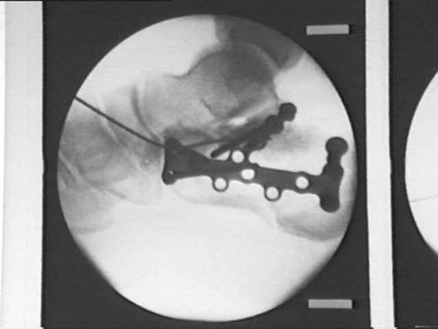
- 3.5 mm reconstruction plate, Synthes 3.5 mm calcaneal plates, or a flattened 3.5 mm 1/3 tubular plate;
- as noted by Carr, et al, in an cadaveric model, the flattened 1/3 tubular plate had a similar load to failure as compared to a 3.5 mm recon plate;
- they concluded that the lower profile 1/3 tubular plate would be a better choice inorder to avoid wound healing complications;
- be aware that bicortical fixation of the medial fragment has a high risk of injurying the FHL, nerves, and vessels;
- direct a 3.5 mm cortical screw or cannulated screw from the subchondral bone of the posterior facet into sustenacular fragment;
- usually these screws must be directed anteriorly inorder to engage the sustentacular fragment;
- more anterior screw needs to be angled upwards;
- posterior screw is angled downwards into dense area of bone;
- use washers if needed;
- these screws will support the fractured articular segments inaddition to securing the sustentacular fragment;
- distally screws are inserted just proximal to the calcaneocuboid joint and are aimed slightly cephalad in order to engage the thick bone underneath the anterior process;
- alterantively consider use of 4.0 cancellous screws rather than bicortical 3.5 mm cortical screws (even for fixation of the medial fragment);
- as pointed out by Bailey et. al., 4.0 mm cancellous screws provided just as good fixation as 3.5 cortical screws since mechanical failure will occur thru ligament disruption rather than screw pullout;
- Hazards:
- be aware that bicortical fixation of the medial fragment has a high risk of injurying the FHL, nerves, and vessels;
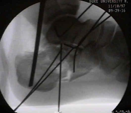
- wires placed in the subchondral bone of posterior facet or anteior to the critical angle of Gissane are at high risk for N/V injuries;
- avoid medial penetration of the posterior facet:
- be aware that the posterior facet slopes from lateral-superior to medial-inferion, and therefore screws which are inserted in a horizontal direction at the superior margin of the posterior facet will penetrate the articular surface;
- in the report by Jordan C, et al (1999), the authors recommend the following:
- at the anterior aspect of the posterior facet, screws need to be angled 5 deg in a plantar direction or need to be placed 1.5 mm below the edge of the articular surface;
- at the mid portion, screws need to be angled 20 deg inferiorly or 10 mm below the joint edge;
- at the posterior portion, screws need to be angled 32 deg inferiorly or 15 mm below the joint edge;
- references:
- Determining the angle of screw placement for internal fixation of calcaneal fractures.
- Internal Fixation of Calcaneus Fractures: An Anatomical Study of Structures at Risk.
Internal fixation of experimental intra-articular calcaneal fractures: A biomechanical analysis of two fixation methods.