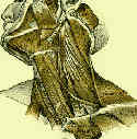
- PreOp Planning:
- consider NG tube and foley catheter;
- have proper sized C-collar available for post operative care;
- position: supine w/ halter cervical traction (3-5 lbs) w/ head turned slightly to the right;
- consider placing a rolled towel between the shoulder blades;
- prep for iliac crest bone graft;
- hall burr is helpful for shaping bone graft;
- Anatomy:
- hyoid cartilage: lies at the level of C3;
- carotid tubercle:
- carotid tubercle is located on the anterolateral aspect of C6;
- longus capitis and anterior scalene muscles attach to it;
- cricoid cartilage: lies at the level of C6;
- vascular structures:
- inferior thyroid artery course horizontally toward midline as branch as branch of thyrocervical trunk at the level of C7;
- carotid sheath (containing carotid artery, internal jugular vein, and the vagus nerve) lie lateral to the dissection;
- vertebral arteries lie posteriorly, and can be injured w/ anterior approach;
- nerves:
- right sided surgical approaches to the cervical spine are generally avoided in order to avoid the aberrant course of the recurrent
laryngeal nerve;
- left recurrent laryngeal nerve, is protected during the dissection as it runs between the trachea and the esophagus;
- cerival sympathetic chain:
- is located anterior to the longus capitis muscle and lateral to the longus coli;
- thoracic duct:
- lies on the left side, lateral to the esophagus;
- at the level of T1, it passes anterior to the anterior scalene muscle, loops around the subclavian artery, and enters the subclavian vein;
- when exposure at this level is required, consider a right sided approach;
- Positioning:
- supine, posterior interscapular role, and Halter traction (5 lbs);

- Surgical Approach:
- transverse incision from the anterior edge of the sternocliedomastoid muscle to a point just shy of the midline;
- incision is carried down to the platysma muscle and fasci;
- metzenbaum scissors are used to bluntly dissect beneath the platysma muscle and are used to elevate
the platysma;
- platysma muscle is incised in line with the incision;
- carotid artery is palpated and protected;
- blunt dissection then procedes posteriorly and medially to the midline of the vertebral body;
- the disc space is identified;
- the anterior edge longus coli muscle is gently mobilized laterally along both sides of the disc space;
- bleeding may be encountered if the dissection proceeds towards the center of the vertebral body;
- Anterior Arthrodesis of Cervical Spine
The anterior cervical approach for traumatic injuries to the cervical spine in children.
The anterior retropharyngeal approach to the upper part of the cervical spine.

