
- See:
- Screw-Plate Angle / Screw Length
- Unstable Intertrochanteric Fractures
- Anatomical Considerations: (see considerations for cannulated screw placement)
- osseous anatomy of proximal femur dictates where internal fixation device should be placed for maximum purchase in femoral head;
- maximum bone density is found in the area where compression & tension trabeculae coalesce in the center of the head;
- in 1838, int. trabecular system of femoral head was described by Ward;
- maximum bone density is found in area where compression & tension trabeculae coalesce in the center of the head;
- when these trabeculae are absent, surgeon can expect higher rate of failure w/ use of device;
- most important aspect of device insertion is secure placement of screw within the proximal fragment;
- hence, insert screw centrally to within 1 cm of the subchondral bone;
- this placement ensures adequate purchase in femoral head & solid fixation of femoral head and neck fragment to shaft fragment;
- central position:
- placement in the center of the femoral head, w/in 5 mm of subchondral bone, results in low rate of clinical failure in pts who have unstable intertrochanteric fracture;
- central, deep screw placement yields the lowest rate of hardware failure, as noted by Baumgaertner, et al (1995);
- as noted by Den Hartog BD, et al (1991), placement of the lag screw in the center of the femoral head (on both radiographic views)
increased the mean load to failure in unstable intertrochanteric hip fractures;
- TAD: sum of the distance from the tip of the screw to the apex of the femoral head on the AP and lateral views.
- as noted by Baumgaertner and Solberg (1997), a TAD of less than 20 mm resulted in a 0% occurance of screw cut out in a study group of 118 fractures;
- The value of the tip-apex distance in predicting failure of fixation of peritrochanteric fractures of the hip.
- Treatment of the unstable intertrochanteric fracture. Effect on the placement of the screw, its angle of insertion, and osteotomy.
- Awareness of tip apex distance reduces failure of fixation of trochanteric fractures of the hip.
- postero-inferior placement:
- inferior placement on AP & slight posterior on lateral x-ray;
- this position may allow impaction & prevent nail from cutting out of head in external rotation and adduction;
- w/ low nail placement, inferior aspect of femoral head is dome shaped and does not allow as deep insertion of the nail;
- advocates of this position note that the calcar is thickest medially & gradually thins as it passes laterally, and therefore there is an
arguement that the best quality of bone is slightly postero-inferior from center;
- as noted by Den Hartog BD, et al, eccentric placement of the lag screw may cause rotatory failure of the fixation;
- references:
- Treatment of the unstable intertrochanteric fracture. Effect on the placement of the screw, its angle of insertion, and osteotomy.
- Predictors of failure for cephalomedullary nailing of proximal femoral fractures
- anterosuperior quadrant:
- bone quality is worst in the anterosuperior quadrant;
- anterosuperior positions are avoided, because bone is weakest in this area, which incr risk of superior cutout of the screw.
- nails in superior aspect of femoral head can inadvertently interrupt lateral epiphyseal vessels that supply most of blood to femoral head;
- Technical Considerations:
- w/ 135 deg plate, insert guide pin approx 2-4 cm below greater trochanter, which is at the level of lesser trochanter (or slightly below it); 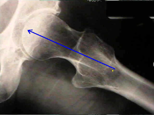
- the guide pin is inserted near the midpoint of the anterior and posterior cortices;
- note that posteromedial comminution may cause the surgon to insert the guide pin too far anteriorly;
- if there is residual retroversion following reduction, then the guide wire should be inserted slightly anterior
to the midpoint so that the wire will not be placed in the anterior half of the femoral head;
- ref: Accuracy of the lesser trochanter for guiding lag screw insertion in hip fracture management.
- use the prominence of the greater trochanter to help determine the proper angle of anteversion;
- in most patients, the greater trochanter will be in line with the femoral neck anteversion;
- ensure that guide pin is well w/in femoral head & lying w/in 5mm of subchondral bone;
- placing guide pin just beneath subchondral bone reduces chance of pin withdrawl during reaming;
- screw should be placed in the central 1/3 of the femoral head between 5-10 mm from subchondral bone;
- there is increase in failure rate if tip of nail is > 12 mm from femoral head subchondral articular surface.
- lag screw length:
- w/ 135 deg plate, typically a 95 mm screw is required;
- determine correct length of lag screw w/ direct measuring gauge;
- this measurement allows for 5 mm of compression
- if more compression is desired, use a shorter screw;
- screw 5 mm shorter permits 10 mm of compression;
- use short-barrel dynamic hip screw sideplate when screw 80 mm or shorter is inserted to maximize available sliding capacity;
- if a shorter screw is used (for better compression) then compression screw must be left in place, so that scew barrel assembly
does not become disengaged;
- if screw sliding brings threads in contact w/ plate barrel, additional impaction will not occur & device becomes biomechanical
equivalent of a rigid nail-plate
Treatment of the unstable intertrochanteric fracture. Effect on the placement of the screw, its angle of insertion and osteotomy.
Subchondral screw fixation for femoral neck fractures
A biomechanical analysis of the sliding hip screw: the question of plate angle.
Trochanteric fractures. Influence of reduction and implant position on impaction and complications.
Awareness of tip apex distance reduces failure of fixation of trochanteric fractures of the hip.
Inadvertent guide wire advancement in hip fracture fixation with fatal outcome
Importance of screw position in intertrochanteric femoral fractures treated by dynamic hip screw
Femoral Head Lag Screw Position for Cephalomedullary Nails: A Biomechanical Analysis
Original Text by Clifford R. Wheeless, III, MD.
Last updated by Clifford R. Wheeless, III, MD on Sunday, April 5, 2015 3:02 pm