
- Discussion:
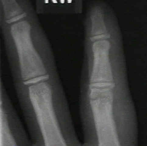
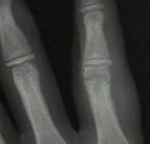
- this fracture most commonly splits off a single condyle, resulting in disruption of the joint and angular deformity of the finger;
- common atheletic injury;
- best diagnosed with an oblique x-ray;
- can be mistreated as sprain which results in finger angulation and irregularity;
- Non Operative Treatment:
- may be indicated if high quality radiographs (including oblique views) fail to show any displacement;
- w/ non operative therapy, regular x-rays need to be taken to ensure that displacement does not occur;
- generally need to immobilize for 3-4 weeks;
- Treatment:
- ORIF is indicated w/ more than 2 mm displacement;
- ORIF requires exact anatomic restoration of articular surface;
- exposure:
- Chamay approach:
- indicated for fractures over the distal 1/3 of the proximal phalanx;
- make a dorsal longitudinal skin incision over the phalanx;
- make a distally based "V shaped" flap incision into the extensor mechanism;
- this allows the central slip to be reflected distally and does not interfere with the lateral bands;
- joint is entered either by splitting the extensor mechanism or by elevating lateral bands and entering the joint dorsolaterally;
- minimize soft tissue stripping and attempt to leave the collateral ligament attached to the condyle;
- stabilize with two K wires (0.28 or 0.35) or a small screw;
- intraosseous wiring is another option;
- bicondylar fractures are more difficult to reduce
Distal unicondylar fractures of the proximal phalanx.