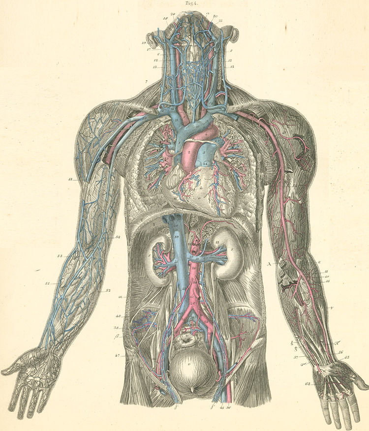
- See: subclavian artery and internal jugular approach

- Anatomy:
- subclavian vein, which in the adult is approximately 3-4 cm long and 1-2 cm in diameter, begins as a continuation of the
axillary vein at the lateral border of the first rib, crosses over the first rib, and passes in front of the anterior scalene muscle;
- anterior scalene muscle is approximately 10-15 mm thick & separates subclavian vein from the subclavian artery which runs
behind the anterior scalene muscle.
- vein continues behind medial third of clavicle where it is immobilized by small attachments to the rib and clavicle;
- at medial border of the anterior scalene muscle and behind sSC joint, the subclavian unites with the IJ to form the innominate, or
brachiocephalic vein;
- large thoracic duct on the left and smaller lymphatic duct on right enter superior margin of subclavian vein near IJ junction;
- Canulation Technique:
- pt to be well hydrated and sedated -
- shoulder blades are thrown back on a towel roll placed beneath spinal column, thereby throwing clavicles backward;
- pts head is neutral and T berg position of 15 deg;
- local infiltration w/ No. 22 needle w/ lidocaine, especially into the periosteum of the clavicle;
- local the left subclavian vein w/ a No. 22 gauge needle;
- have introducer needle present inorder to retrace path;
- ensure that the needle is kept no more than 10 to 15 deg from horizontal;
- subclavian artery, lung, and brachial plexus are all posterior to subclavian vein; if the vein is not cannulated, at least the other structures will not be hit
Prevention of Intravascular Catheter–Related Infections.
Central Venous Catheterization
Implement the Central Line Bundle
Central Venous Catheterization — Subclavian Vein
Effect of Patient Position on Size and Location of the Subclavian Vein for Percutaneous Puncture

