
- Discussion:
 - as with all femur fractures be sure that a femoral neck fracture is not present;
- Operative Technique:
- position:
- supine with several folded sheets placed underneath the injured thigh and knee;
- the injuried thigh must be flexed and elevated higher than the uninjured thigh in order to
obtain a good lateral view;
- incision:
- anterior longitudinal incision from inferior patella to the tibial tubercle;
- patellar paratenon is dissected out as a distinct layer;
- patella tendon is incised longitudinally and is retracted w/ Gelpi retractors;
- ligamentum mucosum is released to allow fat pad to drop inferiorly;
- palpate the PCL tendon;
- percutaneous insertion stratedgy:
- requirements: an adequate closed reduction should be possible (or otherwise an
open reduction strategy will be required);
- reduction:
- first reduce fracture w/ traction, w/ guide pins or K wires, w/ care to avoid screw
placement that would interfere w/ central nail placement;
- alternatively, a large tenaculum clamp can be applied percutaneously to hold the reduction;
- remember that reduction is often achieved with full extension, and temporary pin fixation is
required so that the reduction is not lost when the knee is flexed in preparation for
nail insertion;
- incision:
- make a 5 cm incision over midline of patellar tendon, and split patella tendon and underlying fat pad longitudinally
in line with incision;
- entry point is 3-4 mm anterior to the PCL insertion using a sharp awl;
- ensure that the guide wire is centralized on two flourscopic views w/ respect to the distal fragment (and not the
proximal shaft);
- once distal fragment has been properly reamed, the distal fragment can be reamed if necessary;
- if reaming has remained central in both fragments, reduction should occur as the the nail is driven across the fracture;
- open reduction stratedgy:
- if fracture reduction cannot be achieved, then an open reduction is required, using a anterior midline incision (similar to a
TKR incision);
- entry point:
- depends in part on the curvature of the nail;
- nails w/ a large distal bend will generally be inserted closer to the PCL insertion, where as nails w/ a smaller bow will be
inserted slightly more anteriorly;
- in general, if the starting hole is too anterior, the fracture site will fall into flexion (and the knee will lose extension);
- if the starting hole is sligtly anterior, then make sure to bury the nail slightly deeper than usual as the front edge may
protrude into the femoral trochlea;
- if the nail is inserted too posteriorly, then the frx will fall into extension (and the knee will lose flexion;
- most often the entry site is 6-8 mm anterior to the PCL and slightly medial to the center of the intercondylar groove
- reaming technique:
- with retrograde reaming, it is important to keep a "rolled bump" under the thigh inorder to keep the hip and knee flexed;
- by keeping the hip and knee flexed muscle forces across the thigh are relaxed which facilitates reduction and reamer passage;
- if hip and knee remain extended, there will be displacement of frx site in lateral plane, which may cause eccentric reaming
and subsequent frx malalignment;
- nail passage:
- use of a longer length nail will help centralized the nail in the isthmus (and facilitates reduction);
- note that with distal femoral fractures, the nail must be inserted in a central position, and the fracture must be reduced prior to
nail passage across the frx site (a malreduction will not magically be corrected after the nail is fully seated);
- blocking screw
- references:
- Blocking Screws for the Treatment of Distal Femur Fractures
- Factors Predictive of Blocking Screw Placement in Retrograde Nailing of Distal Femur Fractures.
- proximal interlocking:
- dynamization
- generally the proximal interlocking screws need to be inserted in the AP plane using a free hand technique;
- sizing the screw may be difficult but one can estimate the proper screw length by noting the relative size of the nail in the
femoral canal;
- if a 10 mm nail takes up about 1/3 of the shaft diameter (on the lateral view) then the proper screw length would be about
30 to 32 mm;
- apply an absorbable suture to the screw neck so that the screw is not lost in the soft tissues;
- note that the femoral artery is located medial to the femur throughout its course;
- all branches of the femoral artery cross at a point 4 cm distal to the lesser trochanter;
- hence, keeping the drill bit above the level of the lesser trochanter and avoiding medial aspect of the bone will help
prevent vascular injury;
- similarly, placement of interlocking screws above level of lesser trochanter reduces the risk of injury to branches of the
femoral nerve;
- note that the deep branch of the medial femoral circumflex artery invariably crosses the femur posteriorly at least 10 mm, and
usually 16 to 20 mm, proximal to the superior aspect of the lesser trochanter;
- references:
- Neurologic and vascular structures at risk during anterior-posterior locking of retrograde femoral nails.
- Anatomy of the medial femoral circumflex artery and its surgical implications.
- Retrograde femoral nailing: computed tomography angiogram demonstrates no relative safe zone for prevention of small diameter arterial vascular injury during proximal anteroposterior interlocking.
- Is There an Optimal Proximal Locking Screw Length in Retrograde Intramedullary Femoral Nailing? Can We Stop Measuring for These Screws?
- Complications:
- secondary femoral fracture:
- has been documented in numerous case reports, and apparently occurs as a result of the stress riser created by an IM nail
which ends in the diaphysis;
- ref: Femoral fracture at the proximal end of an intramedullary supracondylar nail: a case report.
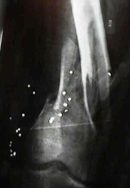
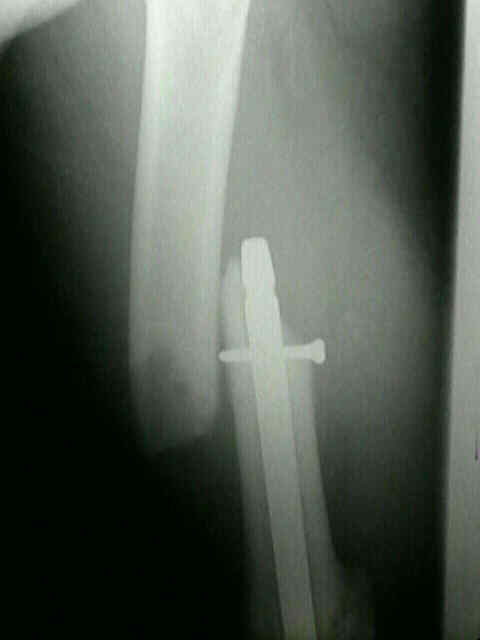
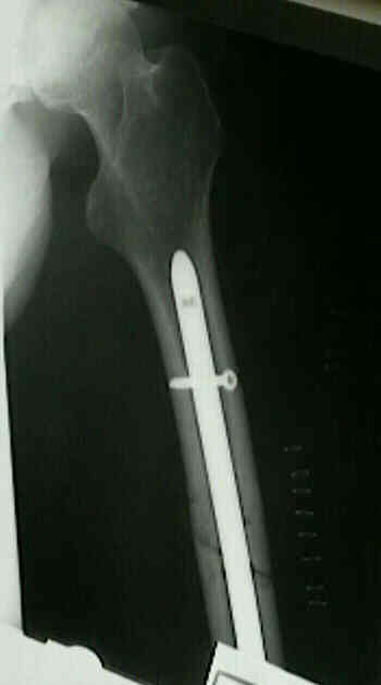
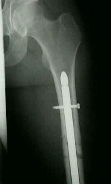
- 24-yr-old male who was initially treated w/ Seligson nail for extra-articular supra-condylar fracture. Apparrently, an attempt
was made to insert a second proximal locking screw (an empty drill hole is seen in the femur);
The fracture occurred 8 months later, at the level of this stress riser;
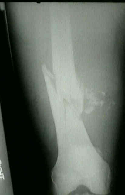
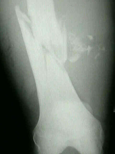
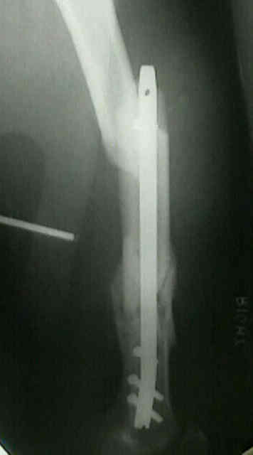

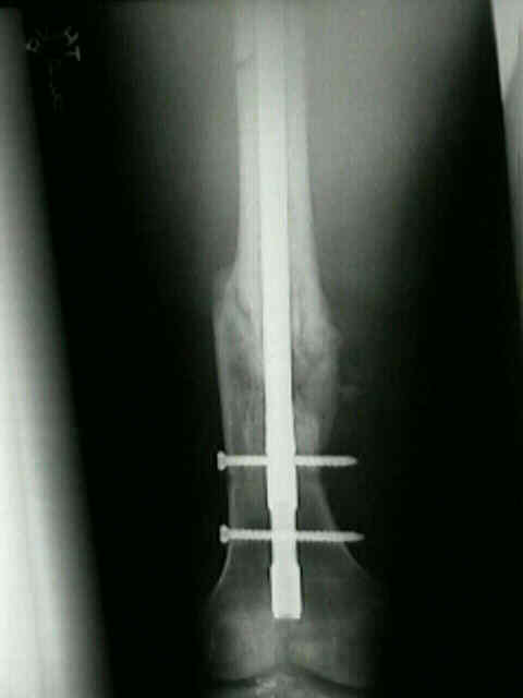
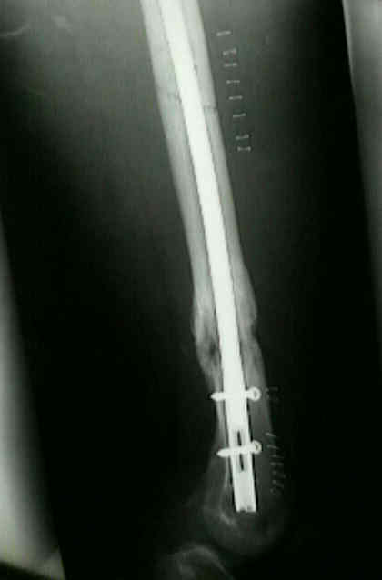
- the previously mentioned supra-condylar fracture which occurred 8 months following insertion of a Seligson Nail,
was treated successively with a retrograde Synthes Femoral Nail
Intramedullary supracondylar nailing of femoral fractures. A preliminary report of the GSH supracondylar nail.
Effects of retrograde femoral intramedullary nailing on the patellofemoral articulation.
Anatomical structures at risk with the proud retrograde femoral nail
The epiphyseal scar as a radiographic landmark for retrograde femoral nail insertion.
Retrograde Versus Antegrade Intramedullary Nailing of Gunshot Diaphyseal Femur Fractures