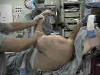
 - Discussion:
- AC joint is situated between the clavicle and acromion;
- acromion has two ossification centers which fuse at age 22 yrs;
- it permits motion in three planes:
- AP gliding of acromion during protraction & retraction of scapula;
- tilting of acromion during abduction & adduction of arm;
- rotation of the clavicle;
- rotation occurs during abduction & adduction of shoulder.
- Anatomy:
- innervation: provided by the suprascapular and lateral pectoral nerves
- ref: The suprascapular nerve and its articular branch to the acromioclavicular joint: an anatomic study
- joint is reinforced by two sets of ligaments:
- AC ligament
- directed horizontally, and functionally the AC joints control horizontal stability;
- palpable shallow depression between end of clavicle & acromion;
- superior AC lig is most important ligament in stabilizing AC joint for normal daily activities;
- coracoclavicular ligaments:
- stronger, vertically directed contains conoid and trapezoid ligaments help to control vertical stability;
- coracoclavicular lig are suspensory ligaments of upper limb;
- conoid:
- is the most important ligament for support of the joint against significant injuries and superior displacement;
- cone shaped which extends between the conoid tubercle on the posterior clavicle and the base of the coracoid;
- trapezoid:
- resists AC joint compression;
- begins anteriorly and laterally to the conoid ligament on the clavicle and inserts on the coracoid process;
- reference: Biomechanical study of the ligamentous system of the acromioclavicular joint.
- Sternoclavicular joint:
- see S.C. joint injury in the adolescent;
- inherently more stable than AC joint; because of this stability & its more protected medial location;
- it is injured less frequently than the acromioclavicular joint.
- Management of Specific Injuries:
- AC joint arthrosis / distal clavicle excision
- The influence of distal clavicle resection and rotator cuff repair on the effectiveness of anterior acromioplasty.
- AC joint septic arthritis:
- Septic arthritis of the acromioclavicular joint - a report of four cases
- Septic arthritis of the acromioclavicular joint.
- Sonographic detection, evaluation and aspiration of infected acromioclavicular joints.
- Primary septic arthritis of the acromio-clavicular joint: case report and review of literature
- AC Joint Separation

- Exam:
- palpate the AC joint during flexion and extension of shoulder;
- distract the arm as it is placed in adduction
- significant prominence of the distal clavicle indicates unstable AC injury;
- BvR test for DJD: resisted shoulder upward flexion with arm hyperadducted;
- ref: Clinical evaluation of acromioclavicular joint pathology: sensitivity of a new test
- Radiology:
- classification of AC separation
- acromioclavicular joint stresses views
- grade I injuries remain nondisplaced;
- type I and type II injuries can be differentiated on stress radiographs;
- w/ pt standing, 10 lb weight is secured to affected upper limb;
- w/ grade II injury, suspended wt displaces AC joint articulation, which increases distance between clavicle & acromion;
- zanca view
- scapular outlet view
- reference: Radiological evaluation of the acromioclavicular joint.
The acromioclavicular joint in rheumatoid arthritis.
Osteolysis of the distal part of the clavicle in male athletes.

