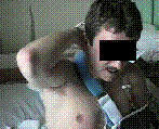
- From Hippocrates to the Eskimo - a history of techniques used to reduce anterior dislocation of the shoulder.
- w/ a witnessed dislocation, consider immediate relocation on field;
- this consists of initial slight abduction and internal derotation of affected arm;
- this can be done without applying a great deal of traction;
- Anesthesia:
- consider intra-articular lidocaine
- give 20 cc of 1% lidocaine or 4 mg/kg (maximum of 200 mg) of 1% lidocaine;
- saves time, and does not require conscious sedation protocol;
- lateral sulcus sign was used to help identify landmarks;
- dislocated humeral head results in a depression just below the acromial edge;
- injection site was 2 cm below either the lateral or posterolateral acromial edge, with the needle directed toward the glenoid cavity;
- references:
- Comparison of intra-articular lignocaine and a suprascapular nerve block for acute anterior shoulder dislocation.
- Intraarticular lidocaine versus intravenous analgesic for reduction of acute anterior shoulder dislocations. A prospective radomized study.
- Anesthetic methods for reduction of acute shoulder dislocations: a prospective randomized study comparing intraarticular lidocaine with intravenous analgesia and sedation..
- Comparison of Intra-Articular Lidocaine and Intravenous Sedation for Reduction of Shoulder Dislocations: a randomized, prospective study
- Milch Technique:

- External Rotation Technique:
- described by Leidelmeyer R., Reduced! A shoulder, subtly and painlessly. Emerg Med 1977;9:233-4.
- success rate of 78%, w/ approx 1% incidence of complication;
- for acute anterior subcoracoid glenohumeral dislocation, however, pts w/ posterior, subglenoid, and subclavicular, or intrathoracic shoulder
dislocations should not undergo this technique;
- avulsion frx of greater tuberosity, or a Hill Sachs lesion, is not a contraindication;
- AP and Transscapular views are obtained;
- r/o neurovascular injury & r/o non displaced humerual neck fractures;
- pts given 5-10 mg of IV valium and morphine;
- w/ pt in supine position, involved arm is slowly & gently adducted against the pt's side;
- elbow is flexed and held at 90 deg;
- physician's other hand holds the pts wrist and slowly externally rotates the forearm w/o longitudinal traction;
- external rotation is extremelly slow, and it is necessary to stop every few degrees until spasm and resistance abates;
- no significant force is required during the external rotation;
- most pts are reduced after about 5 min of exteranl rotation, when forearm reached the coronal plane and was at right angles to body;
- some dislocations reduced after reaching the coronal plane, when the forearm was initially rotated back internally;
- prior to postreduction films, the narcotic antagonist nalaxone is administered IV;
- after reduction is confirmed, neurovascular status is documented;
- Scapular rotational maneuver:
- greater tuberosity is fixed on the anterior glenoid rim;
- as a result, the neck of the scapula is raised and carried medially, which leaves the inferior tip of the scapula in an abducted position
- w/ pt lying prone on the table, the injured side is allowed to hang in dependent position off the edge of the table
- scapula is then manipulated to open the front aspect of the joint so that the congruence of the head and the glenoid is restored,
allowing it to slide back into position;
- prone scaular manipulation was conducted by manually applying downward traction on the affected arm;
- some recommend hanging wts on the flexed or extended arm;
- patient is placed in the prone position on the examining table with shoulder in a position of 90 deg of forward flexion and slight external rotation;
- forearm is suspended from table w/ wrist secured & elbow flexed;
- traction on the forearm is maintained with 5 to 15 lbs for a variable period, usually less than five minutes;
- as pt begins to relax, w/ or w/o aid of sedatives, surgeon pushes on tip of scapula medially (lifting on it occassionally), while
simultaneously rotating the superior aspect of scapula laterally;
- a palpable clunk signals reduction;
- succes with this method often depends on the patient's position on the examing table;
- maintaining a completely flat position is important to avoid excessive tilting, ie. shoulder abduction;
- consider addition of dorsal displacement of the scapula during medial displacement, and consider need for external rotation of the arm;
- external rotation should not be performed with traction;
- traction on the externally rotated humerus elevates the humeral head from the glenoid rim allowing the scapular to be mainpulated back
into anatomic position, effecting reduction of the humeral head;
- ref: Comparison of Intra-Articular Lidocaine and Intravenous Sedation for Reduction of Shoulder Dislocations: a randomized, prospective study
- Seated Scapular Manipulation Technique:
- scapula was manipulated by adduction inferior tip using thumb pressure & stabilizing superior aspect of the scapular w/ cephalad hand;
- dorsal displacement of the tip of the scapula was not included specifically in the description of the technique, although some
naturally occurs with medial displacement of the tip;
- seated scapular manipulation allows the pt to remain seated upright;
- unaffected shoulder is placed firmly against an immobile support such as a wall or the raised head of a stretcher;
- surgeon grasps the wrist of the patients affected side and slowly raises this to the horizontal plane;
- firm but gentle, forward traction is applied with counterbalancing provided by placing the palm of the extended free arm over the
pts mid clavicular region;
- usually only mild traction is required;
- Stimson's technique:
- pt is placed in prone position on the edge of the table while downward pressure is gently applied;
- appropriate weights are taped to the wrist: begin w/ 5 lbs
- this method may take 15 to 20 minutes;
- pt is placed prone on a bed or stretcher with the affected arm and shoulder hanging vertically;
- elbow is positioned in 90 deg;
- while holding patient's wrist with one hand, the surgeon places his other wrist over pt's forearm (near elbow) & graps some
supporting structure underneath mattress, preferably bed frame;
- in this position considerable downward traction can be exerted for prolonged period of time because of the leverage exerted by manipulator's wrist;
- gentle rocking motion may be carried out with the hand holding wrist;
- the key point with this method is that the shoulder must be protracted (w/ the scapula pushed anteriorly)
- this movement increases the anteversion ofo the glenoid cavity, which allows reduction;
- ref: Comparison of Intra-Articular Lidocaine and Intravenous Sedation for Reduction of Shoulder Dislocations: A Randomized, Prospective Study.
- Method of Shoulder Reduction in the Elderly:
- consider applying and monitoring a pulse ox, inorder to monitor the wave form, during the reduction;
- pt is given adequate analgesia & muscle relaxants & is seated in chair;
- usually a combination of valium and demerol is used;
- surgeon stands behind the patient and inserts his flexed forearm into axilla of the affected shoulder;
- his free hand is placed on the flexed forearm of the pt and gentle traction is applied;
- surgeon's forearm pulls in a proximal and lateral direction and lever's the head of the humerus into the socket;
- traction is then released;
- variation of this manuever involves using traction and adduction, w/ fist in the axilla serving as a passive fulcrum
Reduction of anterior shoulder dislocations by scapular manipulation.
The external rotation method for reduction of acute anterior shoulder dislocations.
Reduction of acute shoulder dislocations using the Eskimo technique: a study of 23 consecutive cases.
A modification of the gravity method of reducing anterior shoulder dislocations.
Scapular manipulation for reduction of anterior shoulder dislocations.
The forward elevation maneuver for reduction of anterior dislocations of the shoulder.
A new method of shoulder reduction in the elderly.
Anteroinferior Shoulder Dislocation: an auto-reduction method without analgesia.
Efficacy of the assisted self-reduction technique for acute anterior shoulder dislocation

