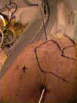
- Technique:
- best visualization of the shoulder joint is through the posterior portal;
- portal usually is located 1.5-3 cm distal & 1-2 cm medial to postero-lateral tip of acromion;
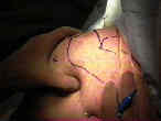
- further refine the proper location of the portal by finding the soft spot, using deep palpation of the thumb along w/ internal and external rotation of the joint;
- the soft spot is formed by the interval between infraspinatus and teres minor;
- always avoid placing the this portal too far laterally, since this will make joint visualization more difficult;
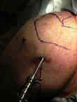
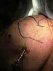
- 18 or 20 gauge spinal needle is then passed into the should joint by aiming just lateral to or at the coracoid process, which is usually well palpated anteriorly;
- note that the scapula is oriented 30 deg anterior to the coronal plane, which is the proper direction of entry;
- inject 5-10 cc of NS w/ epinephrine to distend the joint, and then disengage the syringe and look for fluid exiting from the needle (which confirms proper placement);
- then inject another 20 cc of fluid to fully distend the joint;
- the distended joint will push the humeral head away from the glenoid, which lessens the chance of iatrogenic chondral injury during trochar insertion;
- 30 deg scope with camera attached is locked in place, and the joint distended by the inflow entrance on the scope itself;
- one should then be able to visualize the joint;
- superior to the subscapularis edge is the interval between the subscapularis and supraspinatus;
- this soft spot is where one can and will establish the anterior portal if an operative procedure is warranted;
- arthroscopic acromioplasty:
- when an arthroscopic acromioplasty is planned (along w/ the shoulder arthroscopy) this portal should be placed slightly higher than normal so that the arthroscope lies more parallel to inferior surface of acromion (allowing better visualization for acromioplasty);
- this higher portal also gives a better perspective of the rotator cuff;
- hazards:
- suprascapular nerve:
- injury may occur from passing instruments along the lateral border of the scapular spine;
- axillary nerve:
- this portal is located approx 3-4 cm superior to quadrangular space, which contains axillary nerve & PHCA


