- Discussion:
- dx is made when there is posterior instability on exam and a bone fragment was seen on x-ray;
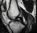
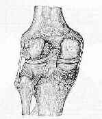
- MRI allows determination of the anatomy of the tear (ie femoral or tibial origin);
- tibial avulsions:
- tibial avulsions are difficult to repair using arthroscopic techniques;
- in the report by Kim SJ, et al (2001), the authors report on methods for managing avulsion fractures of the
posterior cruciate ligament from the tibia;
- 13 patients (14 knees) who had an avulsion frx of the PCL were treated w/ an arthroscopic procedure;
- 11 patients underwent the operation in the acute phase (4 to 10 days after the injury), and 2 patients had delayed surgery (at 19 and 20 months after
the injury) because of nonunion;
- choice of fixation method was based on the size of the avulsed fragment;
- 6 knees that had a small bone fragment (<10 mm) with comminution were fixed with use of multiple sutures;
- 2 knees that had a small bone fragment without comminution were fixed with 23-gauge wires;
- 2 knees that had a medium-sized fragment (10 to 20 mm) were fixed with Kirschner wires;
- 4 knees that had a large single fragment of bone (>20 mm) that involved the condyles were fixed with one or two cannulated screws;
- all patients had osseous union as determined on radiographs;
- 3 injured knees in two patients showed limitation of motion after the operation;
- these patients had been immobilized for two or three months after the surgery because of concomitant fractures.
- 11 patients who had undergone the operation in the acute phase, including two in whom postoperative arthrofibrosis had developed, showed no or
trace posterior instability following the procedure;
- there were better results in patients in whom surgery had been performed in acute phase than in patients in whom operation had been delayed;
- references:
- Arthroscopically Assisted Treatment of Avulsion Fractures of the Posterior Cruciate Ligament from the Tibia
- Treatment of Posterior Cruciate Ligament Tibial Avulsion Fractures Through a Modified Open Posterior Approach: Operative Technique and 12- to 48-Month Outcomes.
- femoral avulsion frx:
- use the PCL guide to effect the reduction of the fracture fragment;
- a small incision is made between the medial femoral epicondyle and the medial border of the patella;
- use a transpatellar portal to grasp the avulsed fragment and oppose it to its femoral bed;
- insert the PCL femoral aiming guide thru the anteromedial portal and apply it against the reduced PCL fragment;
- two guide pins are inserted on either side of the frx fragment;
- sequentially pull the guidewires and in their place, insert a cannulated suture passer in their place;
- as each suture passer enters the joint, an arm of a No 5 ethibond suture is placed into the mouth of the suture passer, and is then drawn out of the joint;
- tension on the sutures will firmly reduce the fracture fragment;
- the sutures are then tied over a bony bridge;
- references:
- Avulsion fractures of the posterior cruciate ligament of the knee. An experimental percutaneous rigid fixation technique under arthroscopic control.
- Isolated avulsion fracture of the tibial attachment of the posterior cruciate ligament.
- Isolated avulsion of the tibial attachment of the posterior cruciate ligament of the knee.
- Arthroscopic posterior cruciate ligament repair
- Primary Repair of Posterior Cruciate Ligament Avulsion Fracture. The Effect of Occult Injury in the Midsubstance on Postoperative Instability.
- Cruciate ligament avulsion fractures.


