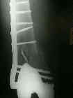
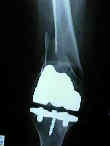
- See: TKR Menu / Supracondylar Femur Frx
- Discussion:
- known risk factors include notching of the femur, osteoporosis, and excessive polyethylene wear (w/ subsequent osteolytic defect)
- other risk factors include osteonecrosis of the femur;
- ref: Morphologic analysis of periprosthetic fractures after hip resurfacing arthroplasty.
- Radiographs:
- often oblique radiographs are needs as well as AP and Lateral views, due to the rotation of the distal fragment;
- lateral radiographs will demonstrate whether the TKR is PCL retaining or sacrificing (the later is more difficult to fix since the intercondylar notch is covered by metal);
- Non Operative Treatment:
- generally non operative treatment is avoided, except in patients with excessive co-morbidity;
- approximately 35% will experience a complication that will require component revision;
- 20% non union rate and 23% rate of malunion;
- permanent knee stiffness is common;
- in the study by Culp et al 1987, one half of patients treated non operatively had increased pain and/or signficant decreased function vs. 13.3% in the control group;
- reference
- Supracondylar fracture of the femur following prosthetic knee arthroplasty
- Retrograde Nailing:
- advantages include ability to exchange the liner (if necessary) and to insert the nail retrograde thru the intercondylar notch - both thru an anterior approach;
- disadvantages
- in the case of a closed box posterior stabilized design retrograde nailing will not be possible;
- even w/ an open box design, the space available for nail passage is limited (usually less than 14 mm), which often means that under-sized nail will have to be used;
- include less than rigid fixation
- blocking screws:
- consider insertion of anterior to posterior blocking screws in both the proximal and distal fracture end (inserted on the non spike end);
- references:
- Extension mal-union of the femoral component after retrograde nailing: no sequelae at six years.
- Fixation of supracondylar frx following total knee arthroplasty: is there any difference comparing angular stable plate fixation vs interlocking nail fixation?
- Fracture Fixation: (see Supracondylar Femur Frx)
- LCP Condylar Plate 4.5/5.0
- see: insertion technique for 95 deg condylar screw
- advantages: allows rigid fixation and early mobilization;
- disadvantages: does not allow for liner replacement and hardware will complicate any future plans for revision;
- if the patient indicates that the TKR was causing pain prior to the fracture, then the need for revision needs to be taken into consideration;
- w/ extensive bone destruction is such that large allograft is needed;
- femoral cortical allograft may be applied to the medial femoral cortex and is secured by a laterally applied plate
The risk of peri-prosthetic fracture after primary and revision total hip and knee replacement
Outcome of periprosthetic distal femoral fractures following knee arthroplasty
Intramedullary Nailing Versus Locked Plate for Treating Supracondylar Periprosthetic Femur Fractures
- Fixation Using Seligson Nail:
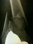
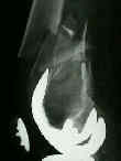
- initial radiographs:
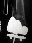
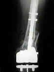
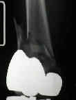 * - 2 weeks postop:
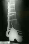
 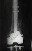 - 6 weeks post op: