- See: Meniscal Tears
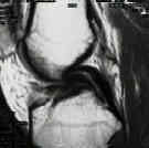
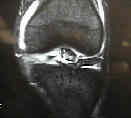
- Discussion:
- vertical longitudinal tear w/ displacement of inner margin
- more common in younger pts & are frequently assoc w/ ACL tear;
- 3 times more common in the medial as compared to the lat compartment
- can produce the classic "lock knee;"
- Clinical Findings:
- in acute cases, the knee may intermittently lock, and will not allow full extension;
- in chronic cases, the bucket handle tear may scar into the intercondylar notch and will defy attempts at reduction;
- strangely enough, some patients will be able to resume near normal atheletic activities despite having a chronic
displaced bucket handle tear;
 - MRI of Knee:
- most commonly missed meniscal tear;
- it may be useful to have the saggital and coronal cuts of the medial compartment up on the x-ray
board at the same time;
- on saggital images, there should be at least two cuts (5 mm cuts) which show the bow tie sign;
- less than 2 boe tie images imply a bucket handle sign;
- "double PCL sign": reliable indicator bucket handle tear;
- Surgical Treatment:
- arthroscopy of the knee
- acute closed reduction:
- attempt to gently extend leg as far as possible, apply valgus, and then begin alternating between internal and external
rotation of knee as well as flexion and extension;
- inspection of medial compartment may be difficult due to displaced meniscus which blocks passage of arthroscope into
that compartment difficult;
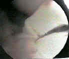
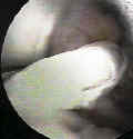
- clear view of the compartment is obtained w/ reduction of fragment, which is performed by using the
"elbow" of the probe to push the meniscus medially;
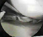 - note the ease of reduction, since this may help determine whether the meniscus should be repaired or
excised;
- meniscal repair:
- indications:
- tear should be within 3-5 mm of the periphery;
- bucket handle should be relatively stable following reduction;
- ref: What are the factors to affect outcome and healing of meniscus bucket handle tears?
- arthroscopic resection:
- attempt to first reduce the meniscus, which will improve visualization;
- if the meniscus is not reducible, excision it can easily be excised in its displaced position;
- approach is to initially detach 95% of either the posterior or anterior rim, followed by complete detachment of the
remaining attachment;
- generally the posterior horn is partially detached prior to complete detachment of the anterior horn;
- obtain maximal visualization and probe the posterior extent of the tear, and then use the probe to determine whether the
excision would be best performed either in front of or posterior to the medial tibial spine;
- use a "curve to the left" cutter for a left knee (and vice versa) inorder to obtain a clean cleavage plan (w/o a remnant portion
of torn meniscus);
- some residents find that they have to cut slightly more posterior than they would have guessed to obtain a clean
meniscal edge;
- leave a small portion of the posterior attachment in intact (usually too large portion is left);
- use a side biter to completely excise the anterior portion (usually one has to cut slightly more anterior than expected);
- an arthroscopic grabber is then firmly clamped onto the displaced meniscus, is locked, and is then "alligator" twisted until the
remnant attachement is broken off;
- a small extension may have to be made in the medial portal inorder to remove the fragment
Bucket-handle tear of the medial meniscus. A case for conservative surgery.
Locked bucket-handle meniscal tears in knees with chronic anterior cruciate ligament deficiency.
Displaced bucket handle tears of the medial meniscus masking anterior cruciate deficiency.

