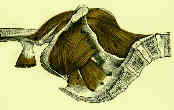
- origin: anterior surface of transverse process, lateral border of vertebral bodies and corresponding
intervertebral discs of T12-L5;
- at the 3 o'clock position, the iliopsoas is composed of 45% tendon and 55% muscle;
- tendon is located directly anterior to the anterior superior capsulolabral complex at the 2-3
o'clock position (hence may cause labral tears)
- insertion: lesser trochanter of femur and for short distance below along medial border of the shaft;
- action: flexion of the thigh at the hip; minimal action in lateral rotatioin and abduction of the thigh;
- synergists: illiacus, adductor brevis, adductor longus, adductor magnus, rectus femoris
- reversed origin insertion action: when the thigh is fixed, the psoas muscle pulls on the vertebrae and flexes the spine
and pelvis on thigh;
- nerve supply: lumbar plexus, L2 > L1, L3, L4; (See innervation)
- references:
- Cross sectional analysis of the iliopsoas tendon and its relationship to the acetabular labrum
- Anatomic Variance of the Iliopsoas Tendon
- Psoas Tendonitis from THR
- ref: Regrowth of the Psoas Tendon After Arthroscopic Tenotomy: A Magnetic Resonance Imaging Study
- Coxa saltans: (snapping hip)
- patients note audible snapping that occurs with flexion and extension of the hip;
- pain usually occurs with activity;
- sub-types:
- external snapping hip
- most common;
- snapping of either the posterior border of the IT band or the anterior border of the gluteus maximus over the
greater trochanter;
- internal snapping hip
- may be a cause of a painful total hip replacement
- clinical findings:
- snapping of the iliopsoas tendon over the iliopectineal eminence occurs as the tendon snaps across bony
prominences when hip is extended from a flexed position;
- patients note painful snapping sensation over the anterior aspect of the groin;
- snapping is reproduced by bringing the hip from a flexed and abducted position to an extended & adducted position;
- snap occurs as the iliopsoas tendon shifting from lateral to medial over the iliopectineal eminence;
- treatment:
- release over pelvic brim vs. lesser trochanter;
- in the report by Gruen GS, et al (2002), the authors discuss surgical treatment of snapping hip syndrome;
- in 30 patients with symptoms in their anterior hip, internal snapping hip was diagnosed by H and P;
- all patients were initially treated nonoperatively and 63% improved and did not require further intervention.
- 11 patients (12 hips) whose symptoms were recalcitrant to PT were offered surgical option of iliopsoas tendon
lengthening;
- procedure was performed via an ilioinguina intrapelvic approach;
- all 11 surgically treated patients (100%) had complete postoperative mitigation of their snapping hip;
- 9 (82%) reported excellent pain relief;
- references:
- The Surgical Treatment of Internal Snapping Hip
- The snapping hip syndrome
- Surgical Correction of the Snapping Iliopsoas Tendon in Adolescents
- Surgical Correction of Internal Coxa Saltans. A 20-Year Consecutive Study.
- Regrowth of the Psoas Tendon After Arthroscopic Tenotomy: A Magnetic Resonance Imaging Study
- Iliopsoas Disorder in Athletes with Groin Pain: Prevalence in 638 Consecutive Patients Assessed with MRI and Clinical Results in 134 Patients with Signal Intensity Changes in the Iliopsoas.
- Psoas Abscess:
- diff dx: septic arthritis of the hip;
- absecesses are experitoneal and follow course of iliopsoas muscle;
- occassionally abscess burrows beneath Poupart's ligament and is seen subQ in the proximal third of the thigh in the adductor
region;
- drainage may be accomplished posteriorly thru Petit's triangle, by a lateral incision along the crest of the ilium, or anteriorly
under Poupart's ligament, depending on the size of the abscess and the area in which it appears
- references: Primary pyogenic abscess of the psoas muscle.
Enlarged iliopsoas bursa. An unusual cause of thigh mass and hip pain.
Primary iliopsoas bursography in the diagnosis of disorders of the hip.
Tendon transfers in the paralytic hip.
The anteromedial approach to the psoas tendon in patients with cerebral palsy

