
CPT David Crawford
MAJ Joanna Branstetter
A. Define the initial evaluation and demonstrate appropriate evaluation techniques of a patient with an extremity vascular injury
I. Initial Evaluation
A. Initial evaluation should always follow acute trauma lifesaving support (ATLS)
B. Application of a tourniquet above the level of injury is appropriate in patients with active hemorrhage who fail to respond to direct compression at the wound
C. Once the patient is stabilized, evaluation of limb perfusion should be performed
D. Palpate all accessible peripheral pulses
1. in the lower limb: brachial, radial, and ulnar artery
2. in the lower limb: femoral, popliteal, dorsalis pedis, and posterior tibial artery
E. If deficits in pulses, exam should progress proximally to identify site of vascular injury
II. Evaluation Techniques
A. Identify “hard” or “soft” signs of vascular injury
1. “hard” signs of vascular injury
a. active pulsatile hemorrhage
b. pulsative or expanding hematoma
c. palpable/audible bruit
d. signs of limb ischemia
e. diminished or absent pulses
2. “soft” signs of vascular injury
a. hypotension or shock
b. developing neurologic deficits
c. stable non-pulsatile hematoma
d. proximity of wound to major vascular structures
B. Presence of peripheral pulses does not rule out vascular injury1
C. Proceed with arterial pressure index if presence of “soft” signs of vascular injury
D. Arterial pressure index is used to compare arterial pressure in an injured compared to a non-injured limb
E. Can be an ankle-brachial index (ABI) for lower extremity trauma or arm-arm index (AAI) for upper extremity trauma
F. To obtain an arterial pressure index, a blood pressure cuff is placed about the ankle or arm of the injured extremity; the cuff is inflated and then slowly deflated and the Doppler probe is used to determine the systolic pressure in the extremity; the probe may be placed over the posterior tibial artery or dorsalis pedis in the lower extremity or the radial or ulner artery in the upper extremity; the systolic pressure in the injured extremity is then compared to the systolic pressure of an uninjured limb (arterial pressure index = Doppler systolic arterial pressure in injured limb/Doppler systolic pressure in uninjured limb)
G. Normal arterial pressure index is greater than 0.9
H. In penetrating trauma, arterial pressure index has a reported sensitivity and specificity of 95% and 97% for significant vascular injury2
I. In knee dislocations, arterial pressure index has a reported sensitivity and specificity of 100% and 100% for significant vascular injury3
B. Understand the indication for vascular stents/shunts/bypass grafts and demonstrate appropriate surgical techniques
I. Patient Preparation
A. Always prep to allow access for proximal vascular control
1. upper extremity: subclavian artery
2. lower extremity: common femoral artery
B. Preparation should include access to uninvolved limb in case vein graft is needed to be harvested
C. Systemic heparinization (50-75 units/kg IV) should be initiated in stable patients with a vascular injury
II. Exposures
A. Longitudinal incisions over named vascular structures allow for widest exposure
B. Curvilinear-type incisions made across joints
C. In contaminated wounds, adequate debridement is essential
D. If ligation of a major artery is required, distal embolectomy should be performed, followed by administration of heparin to preserve collateral circulation4
III. Stents
A. Currently there is a limited role for endovascular procedures in penetrating extremity trauma
B. Endovascular stents may be utilized for blunt trauma or proximal arterial injury where open surgical exposure is difficult (i.e., iliac artery)
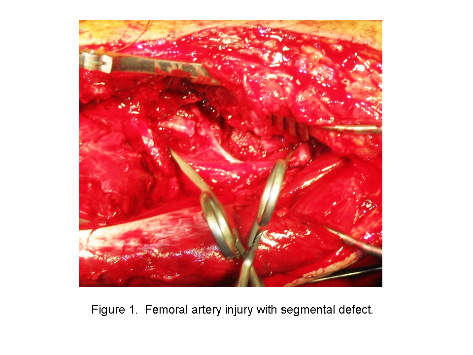
IV. Shunts (see figure 1, figure 2, figure 3)
.png)
.png)
A. Temporary revascularization can be performed with intraluminal shunts
B. After placement, patency should be confirmed with intraoperative continuous wave Doppler
C. Shunts may remain patent for 24 hours without systemic heparinization5
D. Shunts have been reported to maintain limb perfusion up to 48 hours during military operations6
V. Bypass Graft (see figure 4)
.png)
A. Greater saphenous vein is a common conduit utilized for lower extremity trauma
B. Veins should be harvested from the uninvolved limb and be reversed to allow directional flow
C. Bypass grafting should usually be performed at a combat support hospital level
D. Grafting or repair in a grossly contaminated wound should be delayed7
E. Prophylactic fasciotomy should be considered after any revascularization procedure
C. Describe techniques for performing extremity arteriography in an austere environment
I. Equipment Needed8
A. 18- to 20-G butterfly needle
B. Three-way stopcock
C. Two 20- to 30-mL syringes
D. Intravenous contrast
II. Technique (see figure 5)
.png)
A. One syringe is used for aspiration and flushing with heparinized saline solution and the second is used to inject full strength contrast
B. Artery is clamped proximal to the injection site
C. Injection of 15 to 20 mL of contrast into the artery proximal to the suspected injury
D. Fluorsocopy during the injection, if available, or X-ray at the completion of the injection
References
1. Rose SC, Moore EE. Trauma angiography: the use of clinical findings to improve patient selection and case preparation. J Trauma. 1988;28(2):240-245.
2. Johansen K, Lynch K, Paun M, Copass M. Non-invasive vascular tests reliably exclude occult arterial trauma in injured extremities. J Trauma. 1991;1(4):515-519.
3. Mills WJ, Barei DP, McNair P. The value of the ankle-brachial index for diagnosing arterial injury after knee dislocation: a prospective study. J Trauma. 2004;56(6):1261-1265.
4. Gorman JF. Combat arterial trauma. Analysis of 106 limb-threatening injuries. Arch Surg. 1969;123:534-579.
5. Dawson D, Putnam T, Light J. Temporary arterial shunts to maintain limb perfusion after arterial injury: an animal study. J Trauma. 1999;47:64-71.
6. Brounts LR, Wickel D, Arrington ED, Place RJ, Rush RM. The use of a temporary intraluminal shunt to restore lower limb perfusion over a 4,000 mile air evacuation in a special operations military setting: a case report. Clin Med Trauma. 2008;1:5-9.
7. Fox CJ, Gillespie DL, O’Donnell SD, et al. Contemporary management of wartime vascular trauma. J Vasc Surg. 2005;41:638–644.
8. Starnes BW, Beekley AC, Sebesta JA, Andersen CA, Rush RM Jr. Extremity vascular injuries on the battlefield: tips for surgeons deploying to war. J Trauma. 2006 Feb;60(2):432-442.
The view(s) expressed herein are those of the author(s) and do not reflect the official policy or position of Brooke Army Medical Center, the U.S. Army Medical Department, the U.S. Army Office of the Surgeon General, the Department of the Army, Department of Defense or the U.S. Government.
Materials and support for The Disaster Preparedness Toolbox is provided by Lt Col. Ky Kobayashi, MD and Col. Benjamin Kam, MD.