
- see: femoral non unions / osteomyelitis
- three types of infection:
- infection of the soft tissues;
- infection of the fracture site
- infection of the entire marrow cavity:(rare but serious and may require AKA);
- infection usually develops with in 3 weeks of IM nailing;
- look for continuous deep thrombing pain, which is worse at night and persisting beyond usual period of postop discomfort;
- intermittent fever, redness, warmth, tenderness, and marked swelling of the thigh are common;
- Management: Infection following IM nailing:
- once a deep infection is diagnosed - the question is whether to remove nail;
- 4 to 6 months is required for frx healing;
- pt must be protected from septicemia, by thorough debridment of sequestra, infected tissue, open wound care and ATB
- acutely infected IM nails should be irrigated and debridded at the frx site as well as the IM canal;
- consider making a hole in distal medial aspect of femur to allow thorough irrigation of the canal;
- stable intramedullary nail:
- there is no gross motion is present, then leave nail in place if - at debridment of infection;
- nail is usually left in place as long as frx fixation is maintained;
- nail is removed if subsequent x-rays show evidence of bone resorption and lossening of the nail;
- unstable intramedullary nail:
- consider whether to add interlocking screws;
- consider whether to add a larger diameter exchange nail;
- it has been shown that rigid stabilization of frx site is imperative in infected nonunions;
- larger nail may be necessary to compensate for the extra reaming;
- after frx union, which can be expected despite the infection, nail can be removed;
- nail is removed, canal overreamed to remove infected granulation tissue from the canal, canal is irrigated thoroughly, & larger nail is inserted;
- drainage hole in the distal medial aspect of the femur may be created to allow thorough irrigation and drainage of the canal;
- after fracture union, IM nail may then be removed & any residual infection is adressed;
- aggressive infection:
- more extensive infection may require external fixation;
- Examples:
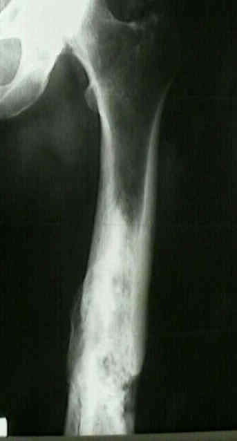
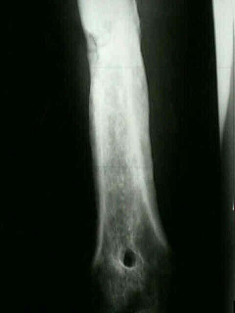
- 35 yo male who developed extensive chronic osteomyelitis following IM nail insertion

- Maintenance of Hardware After Early Postoperative Infection Following Fracture Internal Fixation