
- Discussion:
- often patients will note pain weeks to months after the cast is removed;
- w/ excessive dorsal tilt, will often develop a symptomatic ulnar mid-carpal instability (or carpus adaptive DISI);
- in the report by Taleisnik and Watson (1984), the average amount of dorsal tilt which caused significant symptoms was 23 deg, however, in one case a patient had symptoms w/ 8 deg of volar tilt;
- Colles fracture: does the anatomical result affect the final function?
- Midcarpal instability caused by malunited fractures of the distal radius.
- Exam:
- w/ excessive dorsal tilt, look for:
- symptoms may include tenderness over lunocapitate and triquetrohamate joints;
- a painful audible snap often results from active ulnar deviation w/ forearm pronation;
- some loss of palmar flexion is usually present;
- grip strength is usually decreased by 50%;
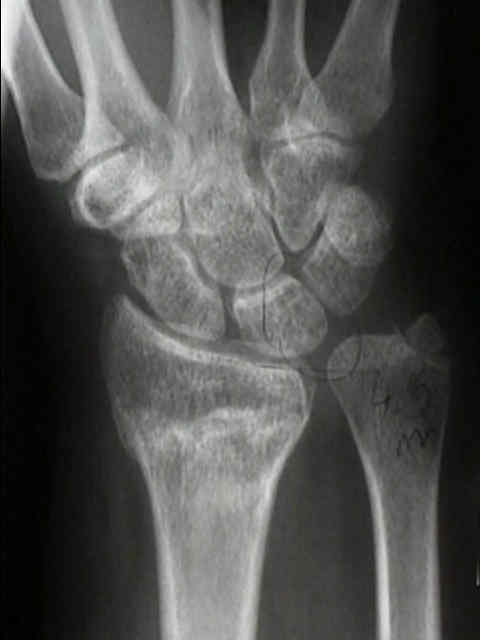
- w/ excessive radial shortening or loss of radial inclination would be more likely to affect the RU joint (limiting pronation and supination);
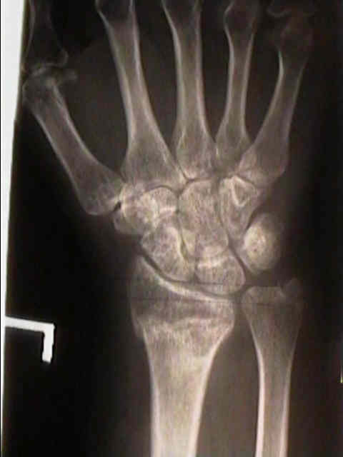
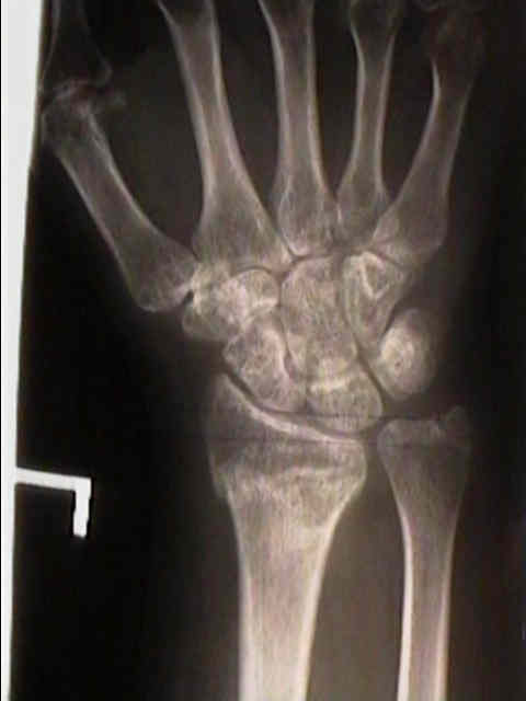
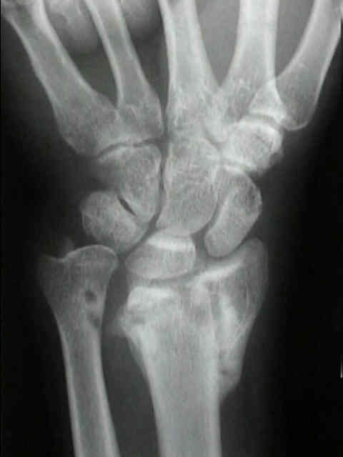
- Radiographs:
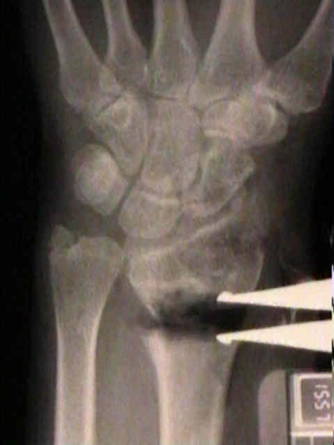
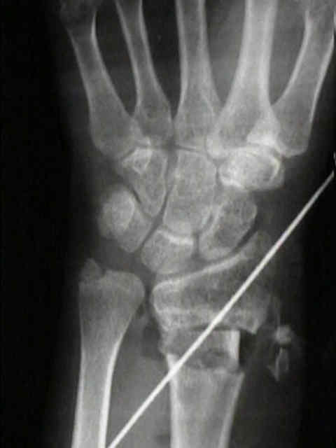
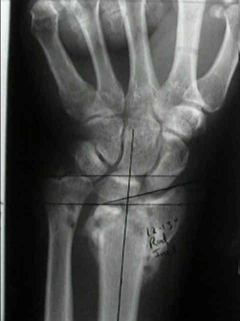
- PA View
- Radial Inclination
- Radial Length
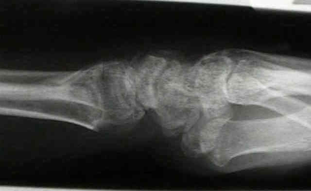
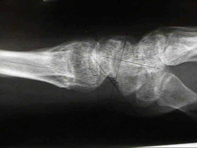
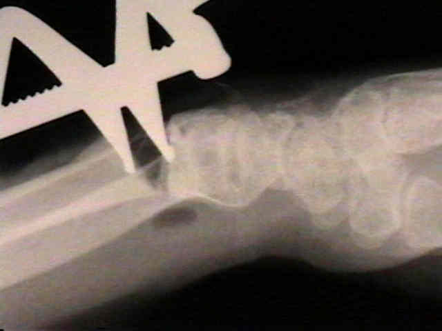
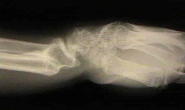
- Lateral View
- Fat Pads (in the case of occult injury)
- Palmar Slope
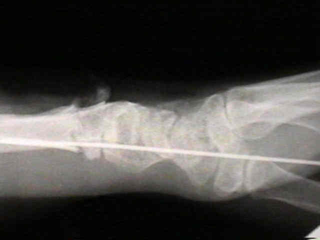
- look for dorsal tilt of the lunate (DISI deformity);
- excessive dorsal tilt is associated w/ ulnar mid carpal instability (or carpus adaptive DISI);
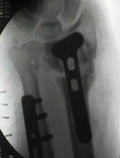
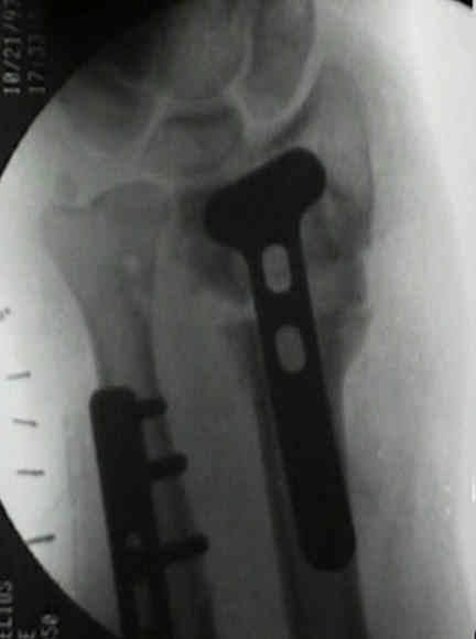
- Opening Wedge Osteotomy:
- preoperative consdierations:
- this procedure is mainly indicated in young active patients;
- ensure that the fracture is fully healed before the osteotomy is performed;
- if the osteotomy is performed before the frx is fully healed, the distal radius may re-fracture as the osteotomy is created;
- radiographs of the opposite wrist should be taken inorder to help judge how much correction is necessary;
- surgical technique:
- dorsal approach to the distal radius;
- distal radius is approached between the 2nd and 4th compartments;
- EPL tendon is mobilized;
- subperiosteal dissection will maximize the amount of soft tissue between the extensor tendons and the plate;
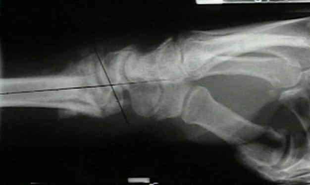
- in the saggital plane, a K wire is inserted perpendicular to the radial surface, at a point several cm proximal to the osteotomy site;
- preparing for the osteotomy:
- a second K wire is inserted just proximal to radial articular surface, at an angle subtended by it and first wire which equals amount of deformity in saggital plane;
- finally a third K wire is inserted parallel to the joint line;
- this ensures that the osteotomy is parallel to the joint line;
- consider using flouro to confirm this, or place a wire thru the joint capsule along the articular surface of the radius;
- Lister's tubercle is removed to produce a more flat surface for the plate;
- osteotomy site is marked 2.5 cm proximal to the wrist joint;
- Homan retractors are inserted to protect the volar soft tissues;
- osteotomy:
- osteotomy is made just proximal to the sigmoid notch;
- in the AP plane, the osteotomy is made at right angles to the radial shaft (as opposed to making it parallel to the radial inclination);
- the later cut may not allow enough room for distal screw fixation;
- in the lateral plane, the osteotomy is made parallel to the dorsal tilt;
- osteotomy is created on dorsal and radial aspects of the distal radius, which allows lengthening and re-creation of volar tilt (against intact volar and ulnar periosteal hinge);
- the osteotomy is spread open w/ laminar spreaders until the K wires are parallel;
- on the radial side of osteotomy, the amount of opening should equal the templated radial length deficit;
- if present, correct any supination deformity of the distal fragment;
- fitting the bone graft:
- laminar spreads hold the osteotomy apart while calipers are used to measure the bony defect;
- radiographs are taken to confirm the correction;
- bone graft is harvested to fit the required dimensions;
- a hall burr can be used to gently shape the bone graft;
- typically the graft will be triangular on the lateral view, and will be trapezoidal on the AP view;
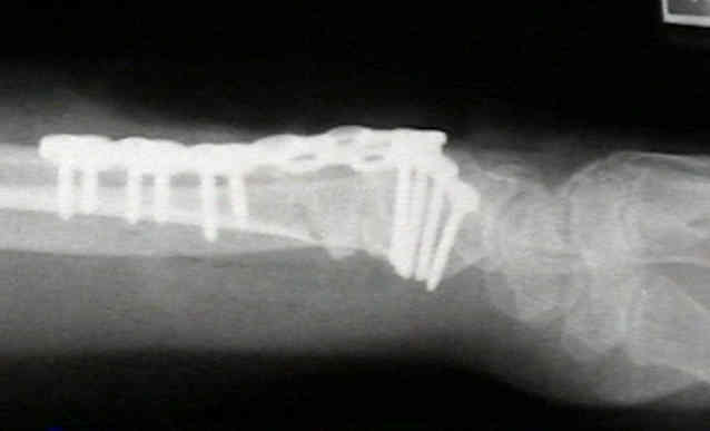
- a plate can be used to secure the graft, but if additional fixation is needed, a lag screw can be inserted from radial styloid to the ulnar cortex of the distal radius;
- assessment of RU joint: (see RU joint)
- these patients will often have an ulnar impaction syndrome;
- following opening wedge osteotomy, check passive supination and pronation;
- w/ a significant deficit, consider Bower's arthroplasty;
- some authors will choose a Darrach procedure


Midcarpal instability caused by malunited fractures of the distal radius.
Opening-wedge osteotomy, bone graft, and external fixation for correction of radius malunion.
Corrective Osteotomy for Intra-Articular Malunion of the Distal Part of the Radius.