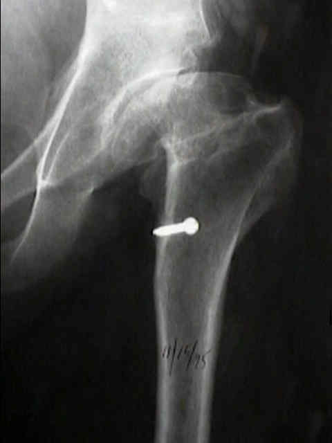
- Femoral Exposure:
- femur is exposed by placing a medium Homan retractor under femoral neck & 2nd under quad. femoris & levering down on fascia lata;
- leg is held in adduction, flexion and internal rotation such that tibia is vertical;
- remove any remaining soft tissue from the posterior and lateral aspect of the neck;
- Positioning:
- place lap sponge into acetabular to collect debris;
- ensure that femur will not move during reaming;
- proximal femur is elevated with jaws or hip skid;
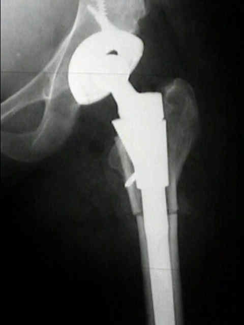
- it is necessary to provide lateral access to femoral canal, because modern femoral stems for cemented use have straight
or nearly straight lateral borders;
- Back Cut:
- most femoral components used today have straight lateral stems or relatively straight stems that necessitate a back cut into the
trochanter, similar to inserting a Moore type prosthesis;
- to provide straight entry into the femoral canal, any remaining lateral bone on the femoral neck and medial cortex of greater trochanter
is removed with box osteotome;
- this can be done with a box osteotome or with a regular chisel;
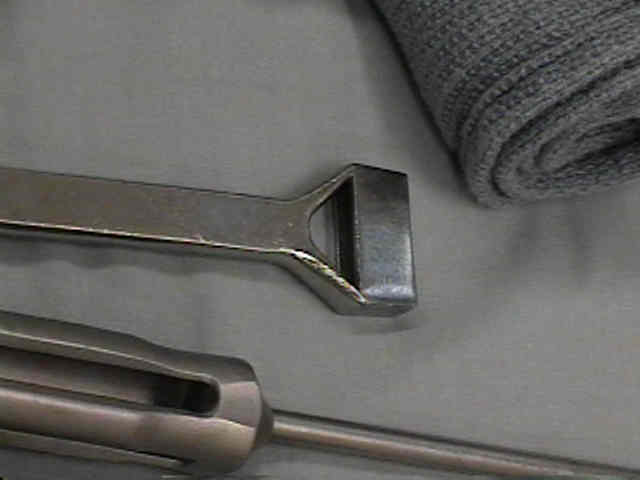
- Enter IM Canal:
- enter IM canal first w/ box osteotome to remove meduallary canal first w/ box osteotome to remove base of femoral neck &
medial aspect of greater trochanter;
- good exposure to meduallary canal is necessary to prevent under-sizing the component and placing it into varus;
- a rasp is then used to enlarge the back cut into the trochanter;
- it is difficult not to overemphasize this back cut;
- if it is not big enough, then varus insertion will occur;
- a high speed burr may help with safe enlargement of the hole;
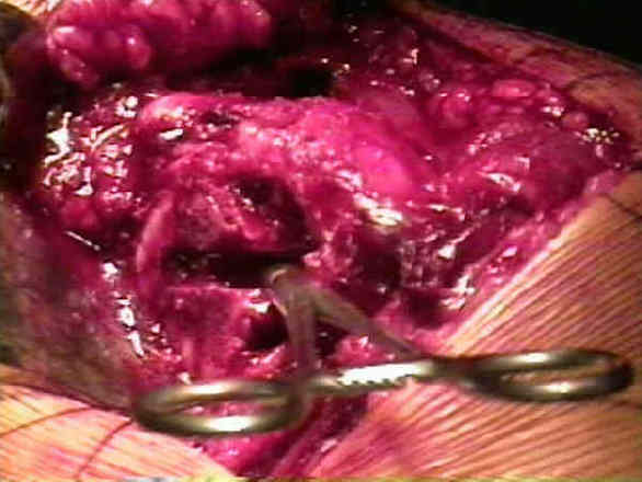
- w/ rongeur or bonx chisel, remove bone at base of neck at its junction w/ greater trochanter so that stem of femoral component will not be
placed in varus;
- femoral component should be placed in slight valgus position;
- w/ hand reamer aim for medial condyle of femur so component will be in slight valgus w/ 5-15 deg of anteversion;
- Misc:
- in some cases, a femoral shaft deformity requires a subtrochanteric osteotomy inorder to allow entry of the stem component into the medullary canal