
- Technique:
- goal is to establish closed continuous drainage thru flexor sheath;
- tourniquet should be used;
- incision & drainage of flexor tendon sheaths are performed from both proximal and distal ends;
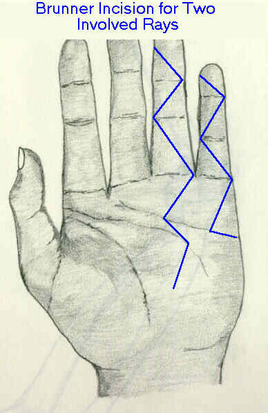
- palmar incision:
- transverse incision is made just proximal to distal palmar crease, over the infected tendon;
- spread thru the palmar aponeurosis;
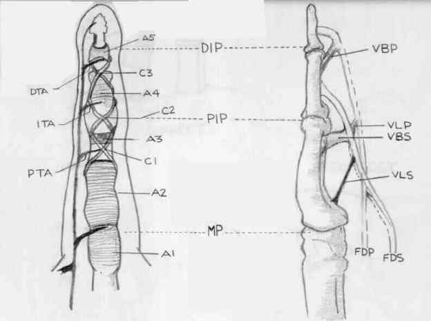
- make incision just proximal to A1 pully and enter into sheath;
- distal incision:
- finger incision may be made either dorslateral at level of middle phalanx or directly on palmar surface at this level;
- incision can also be made in the distal flexor crease of digit;
- distal sheath is exposed thru ulnar midaxial incision & opened;
- enter sheath between annular pulleys, insert small catheter (size no. 5 Fr)
- evaluation of flexor tendons:
- flexor tendon may have to be excised;
- after the infection has been eradicated and the wound closed, consider free tendon grafting and staged tendon reconstruction;
- rebuild pulleys at the time of prosthesis insertion;
- irrigation:
- thread a soft catheter (No. 5 pediatric feeding tube) into distal incision;
- alternatively, 16 ga. polyethylene catheter is inserted into sheath;
- drain is brought out thru skin and the skin is loosely sutured;
- irrigate w/ either sterile saline or sterile Ringer lactate solution;
- sheath is irrigated with 25 to 50 ml of saline/hour;
- antibiotics are not added to the fluid, since this might invite an additional inflammatory reaction in the sheath;
- dressing should contain fluffed gauze and ADB pads to absorb fluid; - dressing is changed as needed