
- Discussion:
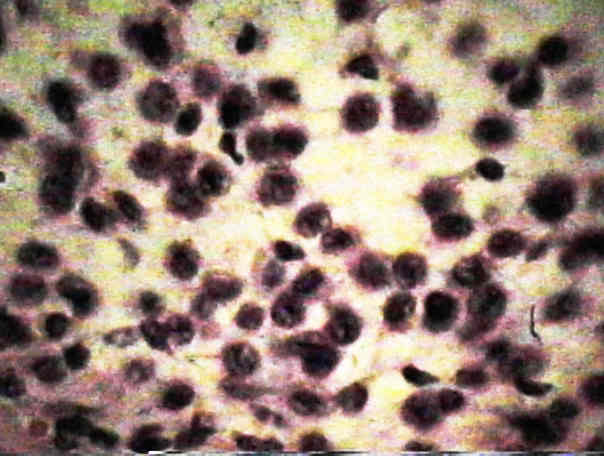
- shows characteristic "cobblestone" pattern;
- areas of round, plump chondroblasts (stones), enmeshed in chondroid matrix;
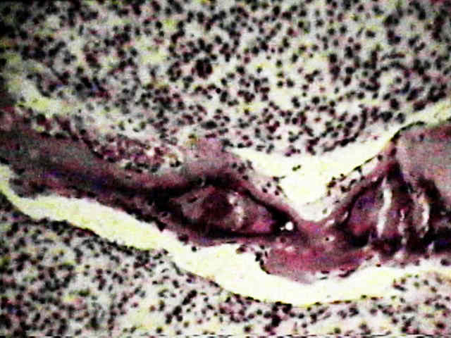
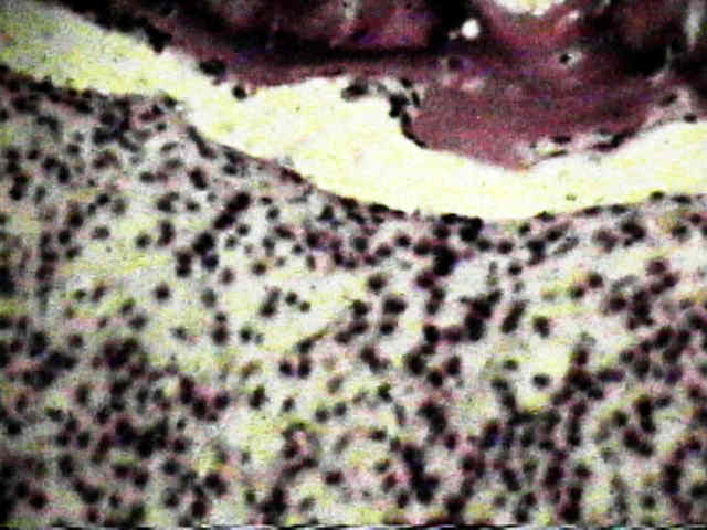
- nodular areas of a chondroid tissue are prominent;
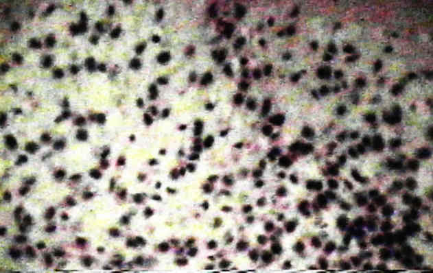
- cartilagenous matrix may have small foci of calcification between between chondroblasts (chicken wire calcification) around islands of cartilage;
- calcification helps to separate chondroblastoma from giant cell tumor;
- cytoplasm: may be pale, and may have an octagonal shape due to abutman against other tumor cells;
- nuclei: are homogenous & oval & contain groove, which resembles EOG;
- secondary Aneurysmal bone cyst
- about 17% of chondroblastomas show features c/w ABC;
- may result in grossly hemorrhagic or cystic specimen;
- clumps of giant cells are found adjacent to areas of spindle cell stroma;
- demonstrates positive S-100 reactivity;
- Diff Dx:
- tuberculosis:
- can cause lytic defects in epiphyseal bone;
- rheumatoid synovitis
- pigmented villonodular synovitis:
- chondrosarcoma:
- may confused w/ chondrosarcoma due to pleomorphic cellular pattern, mitotic figures & multinucleated giant cells between chondroid areas