

- See:
- Excursion, of Tendons
- Staged Flexor Tendon Repair
- Tendon Repair Technique
- Pulley Reconstruction
- Tendon Sheath Anatomy

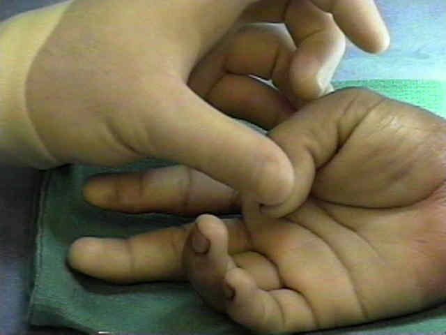
- Anatomy:
- on volar aspect of finger, FDP passes thru FDS to insert on distal phalanx;
- both long flexor tendons are tightly enclosed in common tendon sheath which corresponds to zone II;
- this anatomical proximity explains the development of adhesions between FDS & FDP tendons & digital fibrous sheaths following injury;
- Contraindications to Tendon Grafting:
- neglected tendon lacerations more than 6 weeks old;
- severely contaminated wound;
- loss of tendon substance of greater than 1.5 cm
- loss of the A2 pully, the A4 pulley, or both;
- if wound is contaminated delayed repair (10 days) is possible w/ good results;
- Tecnique:
- graft bed is prepared prior to harvest of the donor tendon;
- remaining portion of the profundus tendon is resected to the mid palm level;
- 1 cm of the distal profundus stump is saved to further augment distal graft anchorage;
- donor tendon:
- donor tendon should not be larger than FDP since this will overcrowd the tendon sheath;
- usual donor tendons include: palmaris longus, extensor indicis, and central toe extensors;
- never use a donor tendon w/ a longitudinal split (such as from the FCR) since this will cause adhesions;
- tendon is passed through the tendon sheath either through the FDS chiasm or around it;
- anchor distal end of tendon graft first (see pull through technique);
- proximal tendon graft anchorage:
- optimizing graft tension
- pulvertaft weave is utilized;
- do not incorporate lumbricals into the repair;
- Post Operative Care
Single-stage flexor tendoplasty in the treatment of flexor tendon injuries
Bridge flexor tendon grafts.
Angiogenesis in healing autogenous flexor-tendon grafts.
Autogenous flexor-tendon grafts. A biomechanical and morphological study in dogs.
Work of flexion after flexor tendon repair according to the placement of sutures.
Original Text by Clifford R. Wheeless, III, MD.
Last updated by Clifford R. Wheeless, III, MD on Thursday, July 9, 2015 2:13 pm