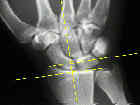
- See:
- AP view
- Carpal height & Carpal height ratio
- Clenched Fist AP
- Radial Inclination
- Radial Length & Width
- Ulnar Variance
- X-ray for Distal Radius Frx
- Discussion:
- AP View is prefered for Wrist Pathology:
- palmar surfaces of most carpal bones are narrower, & these bones are best profiled in AP projection because of the alignment of the peri-articular cortices with the diverging incident x-ray beam;
- position of ulnar styloid will help determine which way film was obtained;
- in standard PA view, ulnar styloid is peripheral;
- in standard AP view, ulnar styloid points centrally;
- radius effectively shortens with pronation and lengthens with supination;
- Technique:
- PA view is taken with the palm flat on the table, elbow abducted to shoulder height and flexed to 90 deg, w/ forearm and wrist in neutral rotation;
- if palm is not flat on the film, CMP joints will be extended and will not be profiled correctly;
- Gilula's arcs:
- on PA view, articular surfaces of carpal bones should be parallel joint spaces should have similar width & have parallel cortical margins;
- broken arc, loss of parallelism, and widening or overlapping of normally parallel joint spaces are strongly suggestive of ligamentous, bone, or joint injury & necessitate further evaluation;
- Radial Ulnar Joints: (PA)
- wrist pronation (PA view) causes positive variance whereas supination achieves the opposite result;
- distal radioulnar joint should measure approximately 2 mm;
- if there is a of a radio-ulnar joint disruption consider CT scan

