
- Discussion:
- although a traumatic etiology is believed to play a major role in production of these lesions, idiopathic osteonecrosis may be another factor;
- anterolateral lesions:
- may result from impaction of talus on fibula as the dorsiflexed ankle is forced into inversion (see ankle sprain);
- the vast majority are caused by trauma;
- these lesions tend to be shallow;
- posteromedial lesions:
- most of these lesions probably arised from trauma (but many will have an atraumatic etiology);
- may result from impaction of posteromedial talus on tibia, as plantar flexion ankle is forced into inversion and exteranal rotation;
- these lesions are deeper and cup shaped;
- exam: palpate just posterior to the medial malleolus with the ankle dorsiflexed (may resemble pending rupture of posterior tib)
- Exam:
- joint line tenderness and effusion;
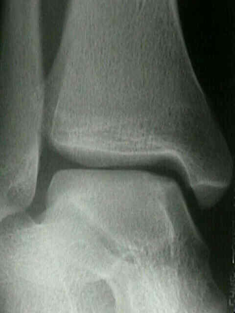
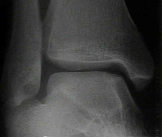
- Radiographs:
- osteochondral frx may be anterior or posterior to dome, requiring plantar or dorsiflexion of ankle to be visible on mortise view;
- if radiographs are negative consider repeat radiographs in 2-4 weeks;
- radiographic classification: (Berndt and Harty)
- note: that radiographic findings may or may not correlate w/ arthroscopic findings nor prognosis;
- I: small area of compression;
- II: partially detached osteochondral lesion;
- III: completely detached, non-displaced fragment;
- IV: detached and displaced fragment;
- Bone Scan:
- usually not ordered until 8-12 weeks following diagnosis;
- a negative bone scan will r/o the diagnosis;
- if bone scan is positive then order either CT or MRI;
- CT Scan:
- offers more accurate staging of the lesion;
- Non Operative Treatment:
- no evidence that non wt bearing cast offers improved results over wt bearing casts;
- no evidence that patients need to be immobilized if they are kept non wt bearing;
- Operative Treatment:
- Arthroscopy of the Ankle:
- osteochondral lesions of the talus can be debrided, and loose bodies and small osteochondral fragments can be removed;
- use of non-invasive or invasive distraction improves access to joint and allows adequate debridement and curettage of bed;
- anterolateral lesions can be addressed from the anterolateral portal;
- in the study by Kumai, et al (1999), the authors noted good clinical results w/ arthroscopy and K wire drilling of the OCD lesions in patients who were younger than 50 years;
- posteromedial lesions can be difficult to access;
- w/ large fragments ORIF may be required, w/ osteotomy of the medial malleolus being required for exposure;
- ORIF allows direct observation of the lesion and accurate repair;
- w/ ORIF immobilization in a non wt bearing cast is required for 6-10 weeks
Osteochondral fractures of the talus. A long-term follow-up.
Osteochondral fractures of the dome of the talus.
Osteochondral lesions of the talus.
Arthroscopic treatment of osteochondral lesions of the talus.
Transchondral fractures (osateochondritis dissecans) of the talus.
Osteochondral lesions of the talus.
Arthroscopic Drilling for the Treatment of Osteochondral Lesions of the Talus