
 - See: Distral Radius Frx Menu
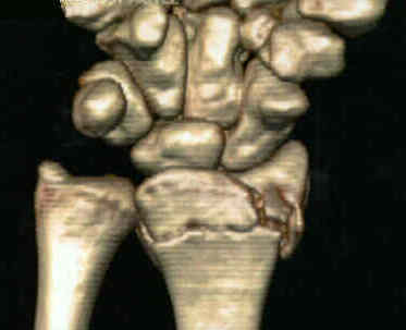
- Dorsal Barton's Frx
- Volar Barton's Frx
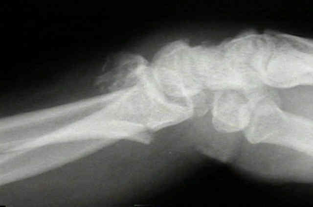
- Unstable Distal Radial Frx
- Discussion:
- mechanism:
- distal radius articular frx principally result from the die punch mechanism of injury that leads to consistent patterns of articular disruption and often w/ radiographic signs of instability (most often DISI deformity);
- anatomy:
- volar surface of distal part of radius is slightly flattened, except for its distal margin, which slopes volarly to form a prominent ridge from which volar radiocarpal ligaments originate.
- short radiolunate ligament originates off of volar margin of lunate facet and attaches to volar surface of lunate and helps maintain stability of the radiolunate articulation;
- reference: Ligament Contribution to Patterns of Articular Fractures of the Distal Radius
- ulnar volar margin of the lunate facet slopes volarly as viewed from proximal to distal and may not be effectively supported by standard implants;
- volar rim of the distal part of the radius is not straight but slopes volarly from radial to ulnar;
- ulnar aspect of the volar rim of the radius is convex distally, forming a palmar prominence of the lunate facet;
- volar lunate facet extends more distally than is expected, which makes it more difficult to get adequate support w/ volar plate fixation;
- fractures of lunate fossa:
- volar shearing fracture with comminution creates a functional radiolunate ligament avulsion, which can lead to instability;
- volar aspect of the lunate fossa bears more load than the scaphoid fossa;
- fractures involving the volar lunate facet articular fragment can therefore be difficult to treat;
- references:
- Loss of Fixation of the Volar Lunate Facet Fragment in Fractures of the Distal Part of the Radius
- The volar extension of the lunate facet of the distal radius: a quantitative anatomic study.
- effects of articular incongruity:
- articular involvement in low-energy frx in older, post-menopausal women has little effect on the generally favorable outcome in these pts;
- in younger patients, these fractures are often result of high energy trauma and may be associated w/ carpal instability, and/or disruption of distal RU joint;
- as noted by Knirk and Jupiter, 91% of wrists that had greater than 1 mm of incongruity developed DJD and 100% of wrists that had more than 2 mm of incongruity develop DJD after an avg 6.7 years;
- as noted by Catalano, et al (1997), residual joint incongruenty will lead to radiographic DJD in about 75% of wrists at an average of 7 years, however, this radiographic appearance does not necessarily correlate with functional status;
- associated injuries:
- TFCC tear
- according to the report by Richards, et al (1997), TFCC tears occurred in 53% of extra-articular distal radius fractures vs 35% of intra-articular fractures;
- scapholunate dissociation:
- as noted by Richards, et al (1997), 21% of patients with intra-articular distal radius frx will have SLD;
- classification:
- frykman classification
- melone classification
- universal classification
- Intra-articular fractures of the distal end of the radius in young adults.
- Displaced intra-articular fractures of the distal aspect of the radius. Long-term results in young adults after open reduction and internal fixation.
- Arthroscopic diagnosis of intra-articular soft tissue injuries associated with distal radial fractures.
- Physical Examimation of Distal Radius Fracture

- Radiographs:
- in the study by Cole, et al (1997), the arc method read off of reformated CT images provided the most reliable method for quantitating articular step off;
- Radiographic evaluation of osseous displacement following intra-articular fractures of the distal radius: reliability of plain radiography versus CT.
- Treatment:
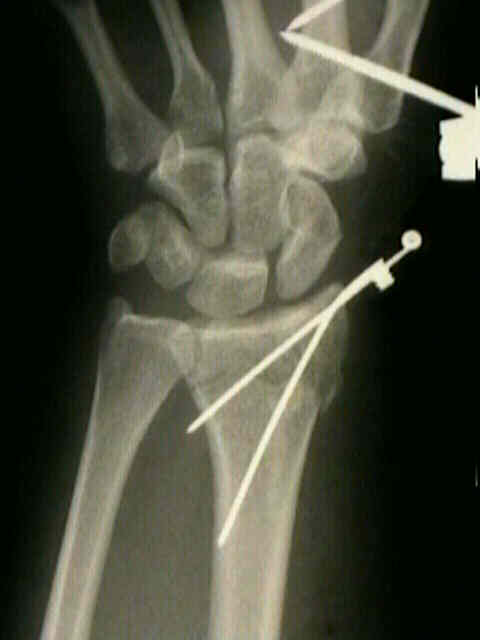
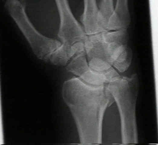
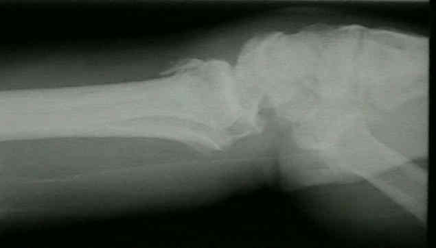
- for patients w/ high energy, shearing, two-part radiocarpal frx-dislocation, (Dorsal or Volar Barton's Frx)
restoration of articular congruity is required to optimize hand and wrist function and prevent post-traumatic arthritis;
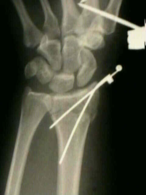
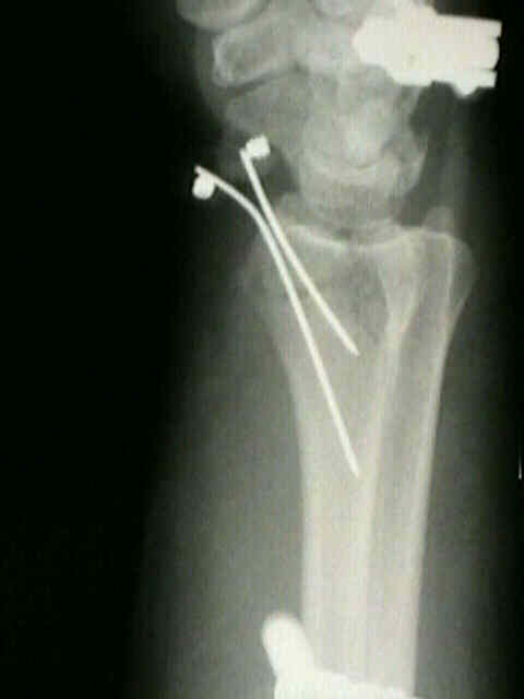
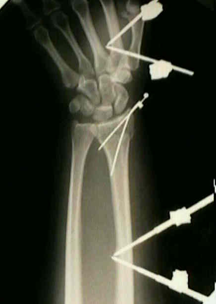
- percutaneous pinning
- external fixation
- references:
- Plaster cast versus external fixation for unstable intraarticular Colles' fractures.
- External fixation for intra-articular fractures of the distal radius.
- The surgical treatment of severe comminuted intraarticular fractures of the distal radius with the small AO external fixation device. A prospective three-and-one-half-year follow-up study.
- ORIF of distal radius fractures
- references:
- The operative treatment of intraarticular fractures of the distal radius.
- Open reduction and internal fixation of comminuted, intraarticular fractures of the distal radius.
- Limited open reduction of the lunate facet in comminuted intra-articular fractures of the distal radius.
- Open reduction and internal fixation of displaced, comminuted intra-articular fractures of the distal end of the radius.
- Use of a Distraction Plate for Distal Radial Fractures with Metaphyseal and Diaphyseal Comminution.
Treatment of displaced articular fractures of the radius.
Displaced intraarticular fractures of the distal radius.
Comminuted intraarticular fractures of the distal radius.
Intra-articular fractures of the distal end of the radius in young adults.
Intraarticular fractures of the distal radius: a cadaveric study to determine if ligamentotaxis restores radiopalmar tilt.
Dynamic external fixation for comminuted intra-articular fractures of the distal end of the radius.
Open treatment for displaced articular fractures of the distal radius.
The Surgical Treatment of Severe Comminuted Intraarticular Fractures of the Distal Radius With the Small AO External Fixation Device: A Prospective Three-and-One-Half-Year Follow-Up Study.
Radiographic evaluation of osseous displacement following intra-articular fractures of the distal radius: reliability of plain radiography versus CT.
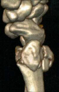
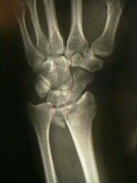
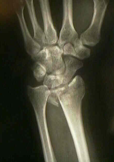
- Case Example:


Original Text by Clifford R. Wheeless, III, MD.
Last updated by on Monday, August 20, 2012 8:14 pm