
- Discussion:
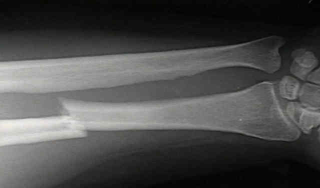
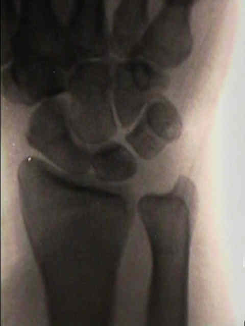
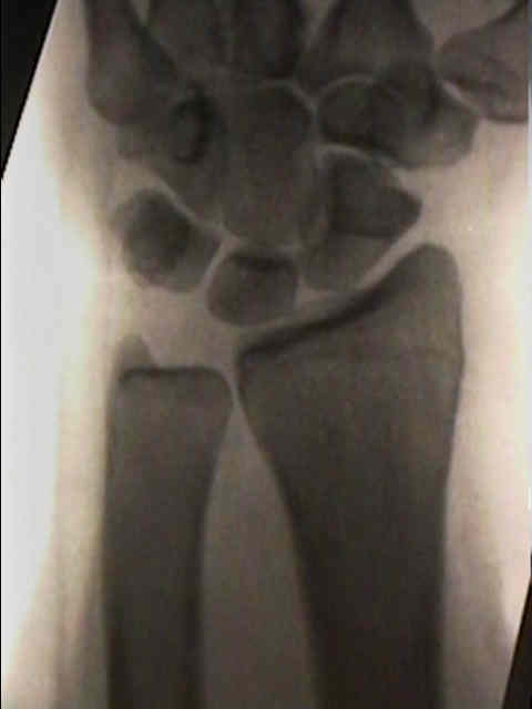
- frx of radial shaft (between middle and distal 1/3's) & dislocation of distal radioulnar joint;
- usually the dislocation is dorsal, but in some cases can be palmar;
- frx is almost always located just above proximal border of pronator quadratus;
- usually there is anterior angulation w/ transverse or short oblique config;
- injury to RU joint may be purely ligamentous (tearing the TFCC), or the ligament complex may remain intact and
the ulnar styloid may be avulsed (in children there may be separation of the distal ulnar epiphysis);
- mechanism: usually direct blows and falls;
- displacing forces:
- wt of hand tends to cause subluxation of distal RU joint & dorsal angulation of the frx radius;
- insertion of pronator quadratus on palmar surface of distal fragment rotates it toward ulna & pulls it in prox & palmar direction;
- brachioradialis causes shortening & rotation of distal RU joint
- Treatment in Children:
- Surgical Treatment in Adults:
- see plating techniques:
- adults tend to have poor results with closed reduction;
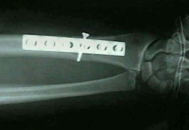
- most adults require compression plates & screws (see below);
- w/ pure transverse frx, 4 hole 4.5 mm plate or 6 hole 3.5 mm plate is acceptable, but if there is comminution a larger plate is necessary;
- no screw should be w/ in 1 cm of frx;
- RU Joint:
- following fixation of the radius, need to reevaluate distal RU joint;
- it is often difficult to evaluate stability of the RU joint w/o opening and directly visualizing the joint;
- even if the supinated joint appears to reduce under flouro, the surgeon's fingers may palpate gross dorsal subluxation;
- in the report by Rettig ME and Raskin KB, the authors categorized these fracture into type I (fractures
within 7.5 cm of midarticular surface of the distal radius) and type II fractures (greater than 7.5 cm from joint surface);
- 22 fractures were type I, and 12 of these cases were associated with intraoperative DRUJ instability;
- 18 type II fractures and were type II, and only one of these frx had intraoperative DRUJ instability after ORIF;
- surgical fixation:
- have the surgical assistant partially supinate the patient's arm
- the surgeon's non dominant hand keeps the joint reduced and helps to "triangulate the k wire" which is driven in with the
surgeon's dominant hand;
- if RU joint is unstable, then K wire fixation is required (K wires are inserted from the ulnar into the radius);
- references:
- Galeazzi fracture-dislocation: a new treatment-oriented classification.
- Surgical Approach:
- Anterior Approach of Henry;
- 5-6 inch longitudinal incision is made, centered over frx in plane between FCR which is retracted ulnarly and BR;
- radial artery is identified & retracted to ulnar side;
- BR & superficial radial nerve are retracted radially;
- frx is located just above proximal border of pronator quadratus;
- insertion of pronator quadratus is freed from radius & reflected ulnarward;
- case example:
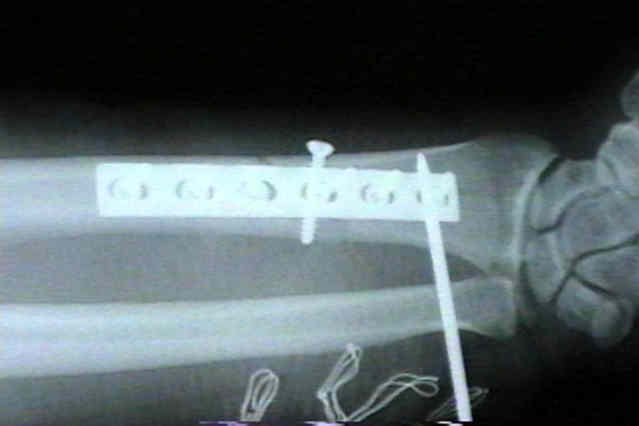
- 30 yo WM who initially underwent ORIF of a Galeazzi frx w/o pin fixation of the RU joint;
- several weeks lateral RU joint diastasis occur which required
closed pinning as a second procedure;


- Post Op
- following ORIF, immobilization in long arm cast with forearm in full supination for 6-8 weeks;
- Complications:
- entrapment of extensor tendons:
- ECU is usually affected but may occur in EDM
- ulnar styloid may sustain avulsion frx & displace into distal
RU joint with the extensor carpi ulnaris tendon.
- exam reveals a vacant ECU sulcus (empty sulcus sign);
- distal radio-ulnar joint is irreducible even after ORIF of radial frx;
- ECU will be found either in RU joint or displaced in an ulnar direction around ulnar head;
- Treatment:
- to avoid chronic instability, the distal radio-ulnar joint is reduced & ECU tendon sheath is repaired;
- surgical repair includes open reduction of distal RU joint, suture repair
of ECU fibro-osseous canal, & ORIF of ulnar styloid frx;
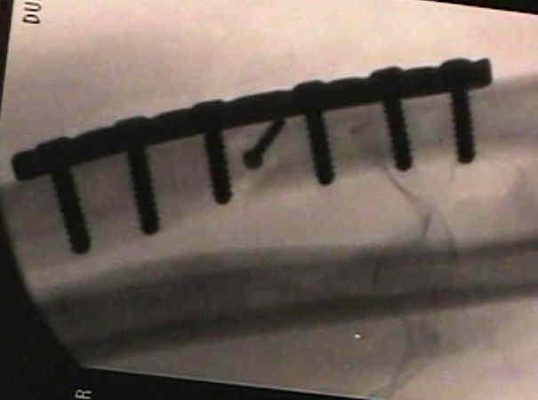
- hardware failure and RU joint subluxation
Results of compression-plating of closed Galeazzi fractures.
Galeazzi injury with an associated fracture of the radial head.
Unstable fracture-dislocations of the forearm (Monteggia and Galeazzi)
Galeazzi fracture-dislocations.
Management of the Galeazzi fracture.
Complex volar distal radioulnar joint dislocation occurring in a Galeazzi fracture.
Internal fixation in 50 cases of Galeazzi fracture.
The Interosseous Membrane of the Forearm: Structure and Its Role in Galeazzi Fractures.
Variant of Galeazzi fracture-dislocation in children.
Galeazzi-equivalent injuries of the wrist in children.